博文
新的显微镜法早期检测抗治疗癌细胞
||
新的显微镜法早期检测抗治疗癌细胞
诸平
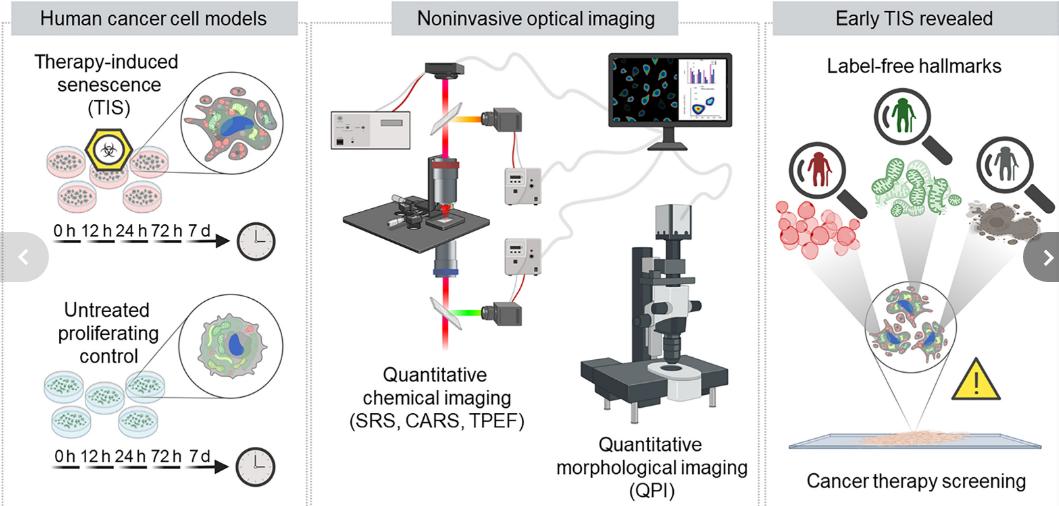
Fig. 1 Schematic illustration of the research design

Fig. 2 Image caption:Ishan Barman
Image Credit: Will Kirk / Johns Hopkins University
据美国约翰霍普金斯大学(Johns Hopkins University)2023年9月22日提供的消息,新的显微镜方法可以早期检测抗治疗癌细胞(New microscopy method detects treatment-resistant cancer cells early),该方法有助于优化临床决策。
化疗是一种对抗癌症的强大武器,但是某些细胞通过进入一种叫做衰老的休眠期(dormant stage called senescence)来抵抗治疗。这些治疗诱导的衰老(therapy-induced senescent 简称TIS)细胞可能对治疗产生耐药性,甚至会变得攻击性和扩散。早期检测TES细胞可能是阻止其生长的关键,但目前的筛查方法在速度和准确性上都有缺陷。
由美国约翰霍普金斯大学(Johns Hopkins University, Baltimore, MD, USA)和意大利米兰理工大学(Politecnico di Milano, Milan, Italy)、意大利国家肿瘤研究院基金会(Fondazione IRCCS Istituto Nazionale Tumori, Milan, Italy)以及意大利国家研究委员会-光子与纳米技术研究所的研究人员组成的研究小组,他们联合开发的新的先进显微镜技术可能会改变这种情况,为临床医生发现这些细胞并调整治疗方案铺平道路。相关研究结果于2023年9月13日已经在《科学进展》(Science Advances) 杂志网站发表——Arianna Bresci, Jeong Hee Kim, Silvia Ghislanzoni, Francesco Manetti, Lintong Wu, Federico Vernuccio, Chiara Ceconello, Salvatore Sorrentino, Ishan Barman, Italia Bongarzone, Giulio Cerullo, Renzo Vanna, Dario Polli. Noninvasive morpho-molecular imaging reveals early therapy-induced senescence in human cancer cells. Science Advances, 13 Sep 2023, Vol 9, Issue 37. DOI: 10.1126/sciadv.adg6231. https://www.science.org/doi/10.1126/sciadv.adg6231
约翰霍普金斯大学怀廷工程学院(Johns Hopkins Whiting School of Engineering)的机械工程副教授伊珊·巴曼(Ishan Barman,Fig. 2)说:“我们的工作证明了改变肿瘤治疗研究的潜力。综合这些显微镜方法可以帮助临床医生做出更明智、更及时的治疗决定。”
这个小组结合了三种尖端的无标签显微镜技术(cutting-edge, label-free microscopy techniques)即相干拉曼散射技术(coherent Raman scattering technique)、多光子吸收技术(multi-photon absorption technique)和光学衍射层析成像(optical diffraction tomography)来观察和分析它们的自然环境中的TIS细胞。与传统的识别方法不同,这些先进的工具使研究人员能够在细胞生命周期内观察和研究细胞的形状、结构以及物理和化学特性。
米兰理工大学物理学副教授达里奥·波利(Dario Polli)说:“重要的是要注意,我们进行这些观察时没有使用侵入性标签(invasive labels),以确保细胞的自然状态得到保存。”
这些技术揭示了TES细胞发生的重大变化。例如,在24小时内,细胞的线粒体,即产生能量的“动力”("powerhouses"),重新排列了自己。在72小时内,这些细胞也开始产生过多的称为脂类(lipids)的脂肪分子,并变得扁平和更大。这项分析使小组能够为这些变化的发展确定明确的时间表。
研究人员说,他们新的快速活筛查技术为未来癌症研究带来了巨大的希望。
“虽然还需要进一步的研究,但我们的研究结果表明,无标签显微镜(label-free microscopy)可以成为提高治疗效果和预防肿瘤复发的重要工具,通过早期检测TIS细胞。这将使临床医生能够在药物导致的衰老使癌症产生耐药性之前调整治疗策略。”意大利国家肿瘤研究所基金会(Fondazione Istituto Nazionale dei Tumori)高级研究助理Italia Bongarzone说。
本研究得到了美国国家普通医学科学研究所(National Institute of General Medical Sciences: DP2GM128198)、美国国家生物医学成像和生物工程研究所(National Institute of Biomedical Imaging and Bioengineering: 2-P41-EB015871)、欧洲联盟项目CRIMSON(European Union project CRIMSON: 101016923)以及伦巴第大区项目NEWMED(Regione Lombardia project NEWMED: POR FESR 2014)的资助。
上述介绍,仅供参考。欲了解更多信息,敬请注意浏览原文或者相关报道。
Anticancer therapy screening in vitro identifies additional treatments and improves clinical outcomes. Systematically, although most tested cells respond to cues with apoptosis, an appreciable portion enters a senescent state, a critical condition potentially driving tumor resistance and relapse. Conventional screening protocols would strongly benefit from prompt identification and monitoring of therapy-induced senescent (TIS) cells in their native form. We combined complementary all-optical, label-free, and quantitative microscopy techniques, based on coherent Raman scattering, multiphoton absorption, and interferometry, to explore the early onset and progression of this phenotype, which has been understudied in unperturbed conditions. We identified TIS manifestations as early as 24 hours following treatment, consisting of substantial mitochondrial rearrangement and increase of volume and dry mass, followed by accumulation of lipid vesicles starting at 72 hours. This work holds the potential to affect anticancer treatment research, by offering a label-free, rapid, and accurate method to identify initial TIS in tumor cells.
https://m.sciencenet.cn/blog-212210-1403719.html
上一篇:小细胞肺癌:克服化学耐药性的新方法
下一篇:大脑炎症标志物终于被破译