博文
2022年11月嘲风作品集(二)
||
▲ Vol 09 Issue 11 | November 01 , 2022
Regulation of iron and cadmium uptake in rice roots by iron(iii) oxide nanoparticles: insights from iron plaque formation, gene expression, and nanoparticle accumulation
Guoyong Huang, Dandan Pan, Milan Wang, Songxiong Zhong, Yingmei Huang, Fangbai Li, Xiaomin Li and Baoshan Xing
The application of iron oxide nanoparticles (FeNPs) can alleviate cadmium (Cd) accumulation in rice. However, the effect of FeNPs on the interaction between Fe and Cd during uptake in rice roots remains poorly understood. Herein, Fe2O3 NPs were applied to rice in a hydroponic experiment under Cd stress. The application of FeNPs significantly decreased the Cd concentrations in roots and shoots and reduced the Fe concentration in shoots. Fe plaque formation was significantly enhanced either by FeNPs or Cd stress; however, the ratios of CdFe plaque/Cdwhole plant and CdFe plaque/FeFe plaque indicated that the contribution of Fe plaque to alleviating Cd uptake was limited. Gene expression quantification suggested that the presence of FeNPs inhibited the uptake of Fe2+ and Cd2+via OsNRAMP5, OsCd1, OsIRT1 and OsIRT2 transporters, but it facilitated the uptake of Fe(III) via the OsYSL15 transporter. TEM-EDS evidenced the accumulation of FeNP aggregates in both the symplast and apoplast of roots, particularly in the symplast, which strongly restricted the root-to-shoot translocation of Cd and Fe, resulting in the Fe accumulation in shoots being even lower than those without FeNPs. This study provides a comprehensive understanding of the regulation mechanisms of Fe and Cd uptake in rice roots by FeNPs from the perspectives of Fe plaque, gene expression, and NP accumulation. The finding that FeNP accumulation in rice roots restricted Fe translocation to the shoot suggested that further investigation needs to optimize the distribution of Fe to rice grains during FeNP application.
https://pubs.rsc.org/en/content/articlelanding/2022/en/d2en00487a
▲ Vol 18 Issue 44 | November 03 , 2022
2D Bismuth@N-Doped Carbon Sheets for Ultrahigh Rate and Stable Potassium Storage
Anding Xu, Qi Zhu, Guilan Li, Caihong Gong, Xue Li, Huaming Chen, Jie Cui, Songping Wu, Zhiguang Xu, Yurong Yan
Potassium-Ion Storage
In article number 2203976, Songping Wu, Zhiguang Xu, Yurong Yan, and co-workers successfully fabricate 2D bismuth@N-doped carbon sheets with a powerful BiOC bond and internal void space for achieving ultrahigh rate and stable potassium-ion storage. The influence of interface microstructure and chemical coupling on the electrochemical properties are revealed by ex/in-situ techniques and density functional theory calculation.
https://onlinelibrary.wiley.com/doi/10.1002/smll.202270235
▲ Vol 10 Issue 41 | November 07 , 2022
Up-conversion charging of a Tb3+-activated garnet phosphor
Tingxing Shi, Feng Liu, Jiahua Zhang and Xiao-Jun Wang
Studies on the up-conversion charging of persistent phosphors are of great academic and practical significance. To date, up-conversion charging designs have been achieved in Cr3+, Mn2+ and Pr3+-activated phosphors. We herein report, for the first time, an up-conversion charging process in the Tb3+-activated garnet phosphor, Gd3Ga5O12:Tb3+, using a 488 nm laser as the excitation source. The delocalized 4f75d state of Tb3+ is excited by absorbing two excitation photons, followed by the charging of the phosphor. Moreover, spectroscopic investigations reveal that an energy-transfer mechanism dominates the up-conversion excitation. This work provides an up-conversion strategy for charging Tb3+-activated phosphors, paving the way toward expanding the scope of persistent phosphors with up-conversion chargeability.
https://pubs.rsc.org/en/content/articlelanding/2022/tc/d2tc03380a
▲ Vol 22 Issue 21 | November 07 , 2022
An integrated microfluidic device for multiplexed imaging of spatial gene expression patterns of Drosophila embryos
Hongcun Zhu, Wenting Shen, Chunxiong Luo and Feng Liu
To reveal the underlying mechanism of the biological function of multicellular systems, it is important to obtain comprehensive spatial gene expression profiles. Among the emerging single-cell spatial-omics techniques, immunofluorescence (IF)-based iterative multiplexed imaging is a promising approach. However, the conventional method is usually costly, time-consuming, labor-intensive, and has low throughput. Moreover, it has yet to be demonstrated in intact multicellular organisms. Here, we developed an integrated microfluidic system to overcome these challenges for quantitatively measuring multiple protein profiles sequentially in situ in the same Drosophila embryo. We designed an array of hydrodynamic trapping sites to automatically capture over ten Drosophila embryos with orientation selectivity at more than 90% trapping rates. We also optimized the geometry of confinement and the on-chip IF protocol to achieve the same high signal-to-noise ratio as the off-chip traditional IF experiments. Moreover, we developed an efficient de-staining protocol by combining on-chip antibody stripping and fluorophore bleaching. Using the same secondary antibody to sequentially stain different genes, we confirmed that the de-stained genes have no detectable interference with the subsequently stained genes, and the gene expression profiles are preserved after multiple cycles of staining and de-staining processes. This preliminary test shows that our newly developed integrated microfluidic system can be a powerful tool for multiplexed imaging of Drosophila embryos. Our work opens a new avenue to design microfluidic chips for multicellular organisms and single-cell spatial-omics techniques.
https://pubs.rsc.org/en/content/articlelanding/2022/lc/d2lc00514j
▲ Vol 41 Issue 06| November 08 , 2022
A chromosome-encoded T4SS independently contributes to horizontal gene transfer in Enterococcus faecalis
Mingxi Hua, Dongfa Dai, Pengcheng Du, Nan Chen, Ang Duan, Jinglin Yue, Hongbing Jia, Chengbo Rong, Ang Li, Hui Zeng, Chen Chen
Bacterial type IV secretion systems (T4SSs) are the specific devices that mediate the dissemination of antibiotic resistant genes via horizontal gene transfer (HGT). Multi-drug-resistant Enterococcus faecalis (E. faecalis) represents a clinical public health threat because of its transferable plasmid with a functional plasmid-encoded (PE)-T4SS. Here, we report a chromosome-encoded (CE)-T4SS that exists in 40% of E. faecalis isolates. Compared with the PE-T4SS, CE-T4SS displays distinct characteristics in protein architecture and is capable of mediating large and genome-wide gene transfer in an imprecise manner. Reciprocal exchange of CE-T4SS- or PE-T4SS-associated origin of transfer (oriT) could disrupt HGT function, indicating that CE-T4SS is an independent system compared with PE-T4SS. Taken together, the CE-T4SS sheds light on the knowledge of HGT in gram-positive bacteria and triggers us to explore more evolutionary mechanisms in E. faecalis.
https://www.sciencedirect.com/science/article/pii/S2211124722014784
▲ Vol 144 Issue 44| November 09 , 2022
Single-Vesicle Infrared Nanoscopy for Noninvasive Tumor Malignancy Diagnosis
Mengfei Xue, Siyuan Ye, Xiaopeng Ma, Fangfu Ye, Chen Wang, Ling Zhu, Yanlian Yang, and Jianing Chen
Protein heterogeneity in molecular expression and structures determines tumorigenesis and is the diagnostic and therapeutic cancer biomarker. Small extracellular vesicles (sEVs) are cell-released nanoscaled membrane-bound vesicles transferring bioactive molecules for intercellular communication and playing essential roles in tumor progression and metastasis. Therefore, protein heterogeneity in tumor-derived sEVs indicates the degree of malignant transformation, providing a noninvasive biomarker for cancer diagnosis and malignancy evaluation. We employ near-field infrared (nano-FTIR) spectroscopy to investigate malignancy-related protein heterogeneity in a single sEV and demonstrate the discriminability of sEV protein heterogeneity to evaluate tumor malignancy and metastasis. We found that the amide I/II adsorption ratio of the sEVs increases with tumor malignancy, the proportion of α-helix + random coil (α-helix and random coil) in sEV proteins decreases with tumor malignancy, and the proportion of β-sheet + β-turn (β-sheet and β-turn) increases with tumor malignancy. These nano-FTIR spectral signatures of the sEVs from the primary tumor tissue of breast cancer patients show high sensitivity and specificity in evaluating tumor metastasis. This study shows the advantages of nano-FTIR in single sEV characterization and demonstrates the significance of sEV protein heterogeneity in cancer diagnosis. It provides a noninvasive solution to elucidate cancer development and facilitates the exploitation of potential cancer biomarkers.
<静远嘲风动漫传媒科技中心>设计制作
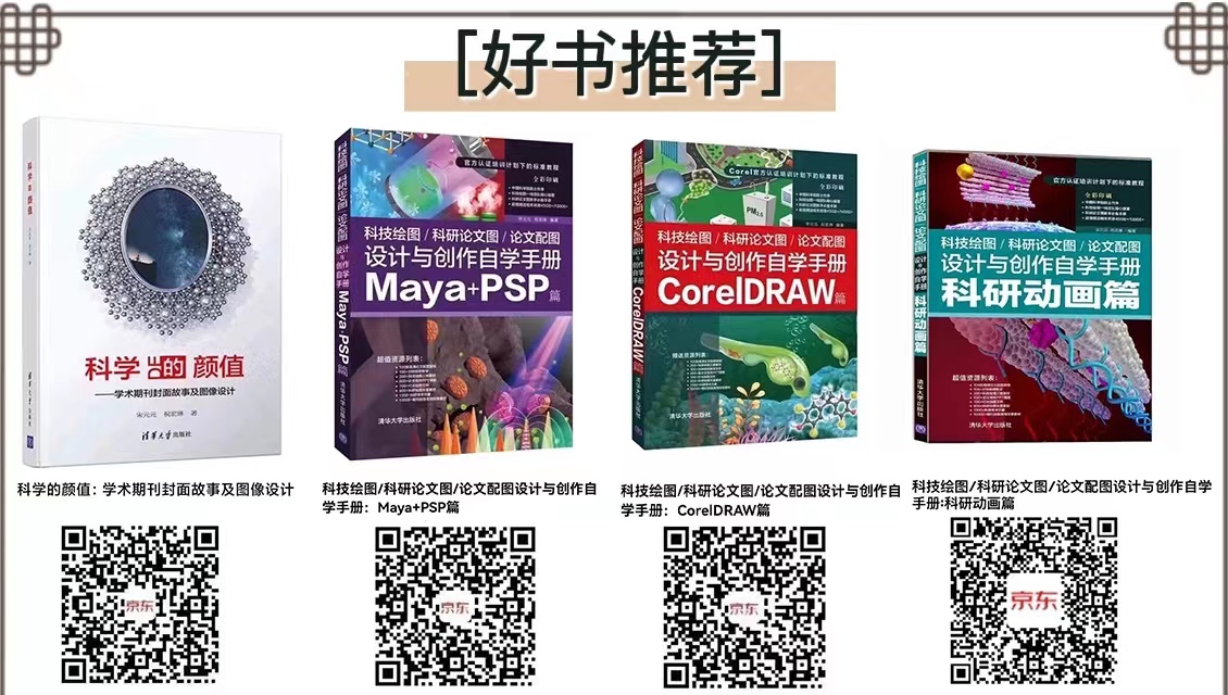
购书链接:
☆科学的颜值:学术期刊封面故事及图像设计
https://item.jd.com/12802188.html
☆科技绘图/科研论文图/论文配图设计与创作自学手册:CorelDRAW篇
https://item.jd.com/13504674.html
☆科技绘图/科研论文图/论文配图设计与创作自学手册:Maya+PSP篇
https://item.jd.com/13504686.html
☆科技绘图/科研论文图/论文配图设计与创作自学手册:科研动画篇
https://item.jd.com/13048467.html#crumb-wrap


静远嘲风(MY Scimage) 成立于2007年,嘲风取自中国传统文化中龙生九子,子子不同的传说,嘲风为守护屋脊之瑞兽,喜登高望远;静远取自成语“宁静致远”,登高莫忘初心,远观而不可务远。


https://m.sciencenet.cn/blog-519111-1374717.html
上一篇:2022年11月嘲风作品集(一)
下一篇:2022年11月嘲风作品集(三)