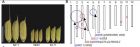
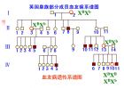
祝贺 Clinical and Translational Medicine (CTM)在2020年度《期刊引证报告》(JCR)中获得首个影响因子—— 7.919 ! 中国目前每年有70万新增肺癌病例,占所有新发癌症的17%。在过去的20年间肺癌的发病率持续上升,五年生存率在16-18%之间。这与缺乏早期诊断和远处转移病灶预警的方法有关。因此,找到与肺癌发生发展相关的精准的生物标志物就显得格外重要。 2020年7月13日, Clinical and Translational Medicine 杂志在线发表了 中国医学科学院肿瘤医院赫捷院士团队 的最新成果 “ Combined detection of aneuploid circulating tumor-derived endothelial cells and circulating tumor cells may improve diagnosis of early stage non-small-cell lung cancer ” 。 许多肿瘤来源的内皮细胞(TECs)落入血液系统成为可循环的TECs(CTECs)。少数情况下循环系统中的非整倍体细胞(非血液系统来源)包括循环系统中的肿瘤细胞(CTCs)和CTECs两种细胞,二者在生物学特性和功能上都不同。CD31是最有代表性的内皮细胞(EC)的标志物之一。因为在循环系统中有大量的正常ECs,所以单独CD31一项指标还不足以检测恶性CTECs。非整倍性8号染色体(CEP8)是识别恶性细胞的重要标志。原位表型和核型联合分析(包括蛋白表达和非整倍性染色体检测)在区分和检测CTCs和CTECs方面有其独特的优势。 图1 通过SE‐\iFISH法检测非小细胞肺癌患者外周血中的CTCs和CTECs 在本项研究中共有98位入选者,其中包括健康志愿者,良性疾病的患者和非小细胞肺癌(NSCLC)早期患者。根据8号染色体的倍数和肿瘤标志物的表达情况对非整倍体CD31+CTECs和CD31-CTCs进行分型,然后用SE-iFISH法对非整倍体CD31+CTECs 和CD31-CTCs的不同亚型进行定量分析。 结果表明CD31-CTCs主要由体积较小(25 μm)的三倍体CTCs和体积较大的超倍体(3五倍体)组成。CD31+CTECs主要由大的超倍体组成。计数CTCs和CTECs的总数对发现恶性结节有较高的敏感性。而三倍体CTCs和CTECs的定量分析对发现恶性结节有很高的特异性。 图2 肺癌发展不同阶段中,总CTC和CTEC数量的变化 结论是在早期NSCLC患者中,同时检测非整倍体CD31+CTECs和CD31-CTCs的特定亚型对发现恶性病灶有十分高的灵敏性和特异性。 总之不同分期的肺腺癌患者,其非整倍体CTCs和CTECs在数量,大小,表面标志物和染色体非整倍性方面都有巨大的差异。非整倍体CTCs和CTECs在肺癌的诊断和治疗中都发挥了各自的作用。未来对非整倍体CTCs和CTECs各亚型的生物学特性进行更加深入的探索将有益于肺癌发病机制和靶向治疗的研究。 参考文献: 1. Chen W, Zheng R, Baade PD, et al. Cancer statistics in China,2015. CA Cancer J Clin . 2016;66(2):115-132. 2. Broncy L, Paterlini-Bréchot P. Clinical impact of circulatingtumor cells in patients with localized prostate cancer. Cells .2019;8(7):676. 3. Pierga JY, Hajage D, Bachelot T, et al. High independent prognostic and predictive value of circulating tumor cells compared with serum tumor markers in a large prospective trial in firstline chemotherapy for metastatic breast cancer patients. AnnOncol . 2012;23(3):618-624. 4. Tinhofer I, Konschak R, Stromberger C, et al. Detection of circulating tumor cells for prediction of recurrence after adjuvantchemoradiation in locally advanced squamous cell carcinoma ofthe head and neck. Ann Oncol . 2014;25(10):2042-2047. 5. Costa C, Muinelo-Romay L, Cebey-López V, et al. Analysis ofa real-world cohort of metastatic breast cancer patients showscirculating tumor cell clusters (CTC-clusters) as predictors ofpatient outcomes. Cancers (Basel).2020;12(5):1111. 6. Lei Y, Sun N, Zhang G, et al. Combined detection of aneuploid circulating tumor-derived endothelial cells and circulatingtumor cells may improve diagnosis of early stage non-small-cell lung cancer. Clin Transl Med . 2020; e128. 原文链接: https://onlinelibrary.wiley.com/doi/full/10.1002/ctm2.128 关于 Clinical and Translational Medicine Clinical and Translational Medicine (CTM)是Wiley出版的英文的开放获取学术期刊。根据科睿唯安2020年6月公布的《期刊引证报告》(JCR),CTM获得的首个影响因子为 7.919 ,在JCR的138本Medicine, Research Experimental 期刊中位列第11,在244本Oncology期刊中位列第27,均处于 Q1 区。 CTM刊登临床和转化医学方面的文章,旨在促进临床前研究向临床应用的转化,加强基础和临床科学家之间的交流。本期刊聚焦新生物技术、生物材料、生物工程、疾病特异性生物标记物、细胞和分子医学、组学科学、生物信息学、应用免疫学、分子成像、药物发现和开发以及监管和卫生政策。CTM竭诚欢迎临床医生、研究人员、决策者和业界人士免费阅读期刊内容并积极向期刊投稿。 撰写:程欣 改编:Tina
选择生儿生女,在我们国家是一种很封建、很落后的想法。我们的先进思想是生男生女都一样。 我是教生物的老师,这里讨论的不是具有先进思想的人类,而是没有思想的动物。不是基于聪明的头脑,而是基于自然选择所塑造的动物的本能,可能从分子、细胞到组织器官甚至行为上的本能。 生儿生女可以选择吗?这样的问题,不同生物学家会有两种不同的思路。 一种思路是大家熟悉的,从动物所具有的分子、细胞、组织器官乃至行为特点,思考研究这一目标是否可能实现,是否存在可能的机制。女性生殖器官是否有能力将仅有一个染色体区别的两类精子(X或Y)区分开并区别对待呢?之前也有些传说,比如酸儿辣女,妈妈喜欢吃酸的,生男孩,喜欢吃辣的,生女孩。可笑的是,这样的传说,换个地区,就反了。西方人 urges would-be moms to eat a more acidic diet if they want to have a girl (建议准妈妈要想生女儿就要多吃酸的食物)。好像准妈妈的饮食影响体液的酸度,体液的酸度使两种(X和Y)精细胞活力出现差别,进而达到选择胎儿性别的目的。这些传说,基本上都是在类似于张悟本粉丝那样的人群中流传,没有任何科学根据,不被科学界认可。除了传说,之前没有任何严肃的科学证据显示动物存在区分两种精细胞的能力。 关于生男生女的另一种思路,是进化生物学家的想法,自然选择有没有可能进化出这种能力。主要讨论具备选择生儿生女能力这一特征的个体是否存在自然选择上的优势,这种突变是否会在种群中扩散开来。这一思路的一个前提是,随机突变,自然界可以出现各种各样稀奇古怪的变异,我们假定这一突变可以并且已经产生了。 1973年,哈佛大学的两位研究人员Robert L. Trivers和Dan E. Willard,从进化生物学的角度,提出动物可能具备选择生儿生女的能力。他们的假说基于一个动物界常见的特征,就是一个雄性动物的精细胞足以为很多很多雌性个体的卵细胞受精。也就是说,一个强壮的雄性个体(如狮王、猴王)所拥有的子女数量远大于一个强壮的雌性个体所能生育的子女个数。如果一位妈妈很强壮,她生出来的子女在下一代的竞争也都占有明显优势,她生儿子会比生女儿拥有更多孙辈后代。反过来,一位妈妈如果很孱弱,生的后代体格在群体中中等偏下,那么她如果生儿子,她的儿子没有机会成为猴王或狮王,她生了儿子和没生孩子一样,她的基因就此打住了、随着她和她儿子的死亡而从地球上消失。如果她选择生女儿,则不一样了。雌性之间竞争远不像雄性那样激烈,她生的女儿还是有机会为她生育外孙、传承接代的。这些权衡不是有意识的权衡,而是自然选择这双无形的巨手在操作。作者也找到了一些动物后代不是雌雄性比1:1的证据。 在我看来,这一假说还是很玄。两种精细胞这样微小的差异,雌性动物体内什么样的机制能够把它们区分开呢?随机突变是可以产生各种各样稀奇古怪的变异,但还有一个前提,就是进化时间足够长,使各种突变有机会出现。区分两种精细胞这样微妙的突变,动物的进化历史有足够长的时间吗?还是运气好,这一突变碰巧了产生了(就像抓彩票一样,所有突变的平均发生频率虽然低,但个别个体还是可能撞上好运的)。因此,上课的时候,我只是把生儿生女的问题作为启发思维的案例讲给学生听,没有把他当成成熟或者可靠的理论讲。 今天看到网络期刊The Scientist的一篇科普文章《 Female Pigs May Sense Sex of Sperm 》“The oviducts of pigs exhibit different gene expression profiles depending on their exposure to sperm with either an X or a Y chromosome, a study shows.”介绍了最近在BMC Genomics上发表的一篇论文C. Alminana et al., “ The battle of the sexes starts in the oviduct: modulation of oviductal transcriptome by X and Y-bearing spermatozoa ,” BMC Genomics, 15:293, 2014. 在哺乳动物中,确实发现了输卵管针对不同性别的精细胞产生了不同的基因表达方式,输卵管的组织免疫体系可能对不同精子产生差别影响。 当然,离确认输卵管能选择不同精细胞还有一定距离,还需要更多证据。但毕竟已经迈出去了第一步。是时候了,我们应该严肃认真对待Trivers和Willard的假说。公奶牛也不产奶,但公黄牛耕地更有力,基础研究不仅是玩玩而已,这方面研究的成果对家畜育种将帮助很大。 顺便鼓励一下年轻的科研人员,你们的思想所受的束缚最少,勇敢地提新想法吧,不管最后证明是对是错,只要根据现有的科学知识,是一个合乎逻辑的想法,就大胆地去发表它。说不清将来哪一天,你的想法启发了他人的实验研究,别人证明了你的假说。如果你想发表生物学假说,我可以帮助选择期刊。
脆性X染色体综合症及其关联的共济失调-上帝偏爱女人的证据 创世纪的时候,上帝按照他自己的样子创造了这个世界上的第一个男人-亚当,后来又觉得亚当孤苦伶仃的样子实在堪怜,于是便取了亚当的一根儿肋骨,制造了夏娃,自此亚当成了夏娃的男人,而夏娃自然就成了亚当的女人! 夏娃听信了蛇的“谣言”,鼓动她的男人偷吃伊甸园里“智慧树”的果子,因此,男人本应该更聪明! 其实,上帝创造亚当,本意是希望他能够做到“难得糊涂”,因为太过聪明会凭空惹来不少麻烦,甚至是杀身之祸,比如三国时期曹操就因为杨修太聪明而把杨修杀无赦...这样男人之间难免会自相残杀... 为了避免这种自相残杀的惨剧发生,也鉴于亚当已经偷吃了那智慧树的果子,上帝于是采取了必要的补救措施-用一条濒临“灭绝”的Y染色体替换掉了亚当体内的一条X染色体,这样使得亚当染色体为XY,而来自亚当肋骨的夏娃依然保留了两条XX染色体,这最终成了男人和女人的内在区别。 上帝把决定人的智慧的基因放在人的X染色体上,并在相应基因的一端插入了一段“垃圾”DNA,这段垃圾DNA就是我们现在已经搞明白了的“CGG”重复序列! 智利中等的人,这段垃圾CGG的重复程度一般比较短,不多于60个重复单位,但很麻烦的是,这段重复序列会像彼诺曹的鼻子那样变长! ( From: Tremor Other Hyperkinet Mov (N Y). 2012; 2 : tre-02-63-375-2. Published online 2012 May 18. ) FMR1 mRNA levels increase with increasing CGG-repeat length (gold segments) throughout the premutation range, and undergo a transition to greatly diminished levels in the full mutation range because of hypermethylation of the FMR1 promoter region. In some instances, methylation mosaicism results in continued production of low-to-moderate levels of mRNA in the full mutation range. RNA toxicity in the premutation range is thought to arise through sequestration, by direct binding to the expanded CGG-repeat element within the FMR1 mRNA, of one or more RNA binding proteins that would normally be associated with other mRNAs. Sequestration in turn leads to loss of the normal function(s) of those proteins, which may include splice modulation and regulation of miRNA production, among other functions. Dysregulation of RNA processing is thought to lead to multiple forms of downstream cellular dysregulation. CCG重复序列的长度超过200个重复单位时,这样的男人会在老年时期出现共济失调---表现为“颤抖”(可能——上帝?),我们临床上称之为“脆性X染色体关联的震颤综合征(Fragile X-associated tremor/ataxia syndrome (FXTAS), 而当这段CGG进一步变长之后,这个男人就会发展为典型的脆性X染色体综合征(马丁-贝尔综合征),这种脆性X染色体综合症直接造成男人的智力低下,这是排在唐式综合征之后,造成男人变傻的第二大原因。 读者诸君看到此时可能会茅塞顿开!上帝控制人类“智慧”能力的秘密就在于此! 他拿掉那男人的一条X染色体,并用一条短的不能在短的Y染色体替换它,同时,他又在直接控制智力的基因处插入了一段CGG重复序列,并使这段重复序列“莫名其妙”地变长或变短! 当这段重复序列很短时,那男人的智力会高些... 当这段重复序列变到一定长度,那男人依然是聪明的,但需要表现出“颤抖”--震颤... 当这段重复序列变得更长之后,那男人无需再颤抖,而是失去了大部分的智力, 便成傻子! 难道这一切都“报应”给男人?而不“报应”女人? 确实是这样,因为女人有两条X染色体,比男人多一倍的机会被免于惩罚! 但是,这样的女人只是“缓刑”,她会有50%的可能性使她生的儿子变成Fragile X-associated tremor/ataxia syndrome (FXTAS)或脆性X染色体综合征! 很麻烦的是,CGG这种长短的改变毫无预兆!因为它并不遵守“蒙德尔遗传”(必然性),而是一种“动态”遗传,这里面存在在“偶然性”! 直到目前为止,此类问题依然得不到解决!已经成为世界性的重大难题之一。 非常不幸!老夫在相关领域干了17年!目前虽然没有变傻,但几乎变疯! 因此不得不祷告: 上帝啊!请您饶恕! Fragile X-associated tremor/ataxia syndrome (FXTAS) 脆性X相关的震颤/共济失调综合征( FXTAS ) 是一种尚未给以足够认识的疾病( a significant cause of late-adult-onset ataxia)。 The etiology is expansion of a trinucleotide repeat to the premutation range (55到200 CGG repeats) in the fragile X mental retardation 1 (FMR1) gene. 该基因扩增 200 CGGs 导致脆性X染色体综合症( fragile X syndrome) 是一种最为常见的可遗传性认知障碍和自闭症的发病原因(The most common heritable cause of cognitive impairment and autism) . Core features of FXTAS include progressive action tremor and gait ataxia; with frequent, more variable features of cognitive decline, especially executive dysfunction, parkinsonism, neuropathy, and autonomic dysfunction. MR imaging shows generalized atrophy and frequently abnormal signal in the middle cerebellar peduncles. Autopsy reveals intranuclear inclusions in neurons and astrocytes and dystrophic white matter. FXTAS is likely due to an RNA toxic gain-of-function of the expanded-repeat mRNA. The disorder typically affects male premutation carriers over age 50, and, less often, females. Females also are at increased risk for primary ovarian insufficiency, chronic muscle pain, and thyroid disease. Treatment targets specific symptoms, but progression of disability is relentless. Although the contribution of FXTAS to the morbidity and mortality of the aging population requires further study, the disorder is likely the most common single-gene form of tremor and ataxia in the older adult population.
染色体做图程序 #hypoloc ==11]; #hyperloc ==11),] # prepareGenomePlot example # construct genomic positions # Chrompos returns a matrix with the positions of the elements on the plot # You can use all kinds of base graphics functions to annotate the chromosomes #png(chroidem.png) setwd(/home/gsc/houston/upload/rheumatology/scleroderma/chromplot/chromeplot) data-read.table(chrome,head=T,sep=\t,as.is=F) hyperloc=data hypoloc=data postscript(chrosomeplot.eps) par(mar=c(0.1,1,1,1)) library(quantsmooth) CHR-hyperloc # Chromosomes MapInfo-hyperloc # position on chromosome CHR-hypoloc # Chromosomes MapInfo-hypoloc # position on chromosome chrompos2-prepareGenomePlot(data.frame(CHR,MapInfo),paintCytobands = TRUE, organism=hsa,sexChromosomes = TRUE,units=bases,cols=green) legend(topright,legend=c(CNV Gain,CNV Loss), col=c(red,green),bty=n,pch=|) points(chrompos1 ,chrompos1 +0.2,pch=|,col=red) points(chrompos2 ,chrompos2 +0.2,pch=|,col=green) title(main=Figure 1 Distribution of the CNV in human systemic scelerosis,line=0.5) dev.off()
p arm of a chromosome: The short arm of a chromosome. The p comes from the French petit meaning small. The letter q was selected to signify the long arm merely because q is the next letter in the alphabet. 也就是说,“p”代表短臂是因为法语单词petit的缩写,而长臂用“q”表示仅仅是因为与短臂相对应,Q紧挨着字母P。
图片来源 http://aobblog.com/2013/01/evolution-of-genome-size-in-carex/ 薹草属包含约 2000 个物种,染色体数目变异较大,并具有全着丝粒染色体。除了少数物种(约 2% )的基因组大小被报道以外,其余仍不得而知;基因组碱基构成( AT/GC )更一无所知。 Lipnerová et al . 报道了 薹草属( Carex ) 157 个物种的基因组大小,变异范围在 0.24-1.64 pg/C 之间,基因组相对较小; GC 含量变异范围在 34.8%-40.6% 之间,相比较其他被子植物而言, GC 含量相对较低。在该属的核心类群发现了 7 个多倍体物种。该属非多倍体物种的基因组大小与染色体数目呈强烈的负相关。作者认为, 频繁的染色体融合或分裂 , 很少的多倍化以及普遍的重复序列增殖或去除 ,是薹草属基因组与核型进化的动力。 原文: Ivana Lipnerová , Petr Bureš , Lucie Horová , and Petr Šmarda Evolution of genome size in Carex (Cyperaceae) in relation to chromosome number and genomic base composition Ann Bot ( 2013 ) 111 ( 1 ): 79 - 94 first published online November 21, 2012 doi: 10.1093/aob/mcs239 http://aob.oxfordjournals.org/content/111/1/79
The objective of this study was to locate chromosomes for improving water and phosphorus-deficiency tolerance of wheat at the seedling stage. A set of Chinese Spring-Egyptian Red wheat substitution lines and their parent Chinese Spring (recipient) and Egyptian Red (donor) cultivars were measured to determine the chromosomal locations of genes controlling water use efficiency (WUE) and phosphorus use efficiency (PUE) under different water and phosphorus conditions. The results underlined that chromosomes 1A, 7A, 7B, and 3A showed higher leaf water use efficiency (WUE(l) = Pn/Tr; Pn = photosynthetic rate; Tr = transpiration rate) under W-P (Hoagland solution with 1/2P), -W-P (Hoagland solution with 1/2P and 10% PEG). Chromosomes 7A, 3D, 2B, 3B, and 4B may carry genes for positive effects on individual plant water use efficiency (WUE(p) = biomass/TWC; TWC = total water consumption) under WP (Hoagland solution), W-P and -W-P treatment. Chromosomes 7A and 7D carry genes for PUE enhancement under WP, -WP (Hoagland solution with 10% PEG) and W-P treatment. Chromosome 7A possibly has genes for controlling WUE and PUE simultaneously, which indicates that WUE and PUE may share the same genetic background. Phenotypic and genetic analysis of the investigated traits showed that photosynthetic rate (Pn) and transpiration rate (Tr), Tr and WUE(l) showed significant positive and negative correlations under WP, W-P, -WP and -W-P, W-P, -WP treatments, respectively. Dry mass (DM), WUE(P), PUT (phosphorus uptake) all showed significant positive correlation under WP, W-P and -WP treatment. PUE and phosphorus uptake (PUT = P uptake per plant) showed significant negative correlation under the four treatments. The results might provide useful information for improving WUE and PUE in wheat genetics. from: www.mdpi.com/1422-0067/10/9/4116/pdf
基因组测序技术和染色体异常鉴定技术同时用于筛查癌症 http://www.biodiscover.com/news/hot/research/103017.html 发布:2012/11/30 来自:中国科学报 阅读数: 1006 导读 约翰霍普金斯大学研究人员将血液基因组测序和DNA测试技术结合后检测癌症用于筛查癌症,监测癌症病人的复发和手术后残留的癌细胞。相关研究成果发表在11月28日的《Science Translational Medicine》杂志上。 在肿瘤初期时,人们能从简单、精度高的血液检查中发现什么?现在,通过排序肿瘤释放在患者血流内的异常DNA,研究人员朝用常规血液检查检测癌症迈出了重要一步。 基因组测序 技术和染色体异常鉴定技术同时用于 筛查癌症 通常,肿瘤DNA检测依靠寻找已知的癌症基因突变来区分癌症DNA和正常DNA。《科学》杂志在线报道称,为了寻找一种不依赖于已知癌症基因组成而检查肿瘤DNA的方法,美国约翰斯•霍普金斯大学医学院Victor Velculescu Luis Diaz实验室的Rebecca Leary和同事,以及来自其他研究机构的合作者,研究了他们手头的观察报告,发现无论哪种类型的癌症,肿瘤细胞总是发生染色体变化。而这暗示着一种能够检测患者血液内染色体异常的检验方法,可能成为一种常规癌症检验法。 首先,研究人员从取自10位结肠癌和乳腺癌患者的血液标本里分离出DNA。然后,他们使用下一代DNA测序技术读取了血液DNA中的全部基因组。结果显示,癌症患者的血液DNA都出现了染色体变化,而10个健康对照组成员的DNA没有异常。相关研究成果刊登于最近的《科学—转化医学》上。 现在,研究人员对新的技术充满希望。“该方法有多种用途。”Velculescu说,例如该技术还能在不进行活组织检查的前提下,判断一位患者应该服用何种药物。他的研究小组致力于追踪肿瘤是否对治疗有响应及手术后是否复发。 不过,目前这种技术并不便宜也不快速。研究中,在每10位患者的相关检测中,仅仅DNA测序就花费数千美元,另外,包括分析在内共耗费一个月的时间。而且,早期癌症检查仍然十分遥远。该技术必须通过筛检大量DNA来发现与肿瘤相关的变化,不过癌症患者血液中来自肿瘤的DNA所占比例从1.4%到47.9%,差异很大。 这种新检测法还可能需要从肿瘤DNA低于0.1%的血液样本中检查出小的、可治愈的肿瘤。研究人员则表示,只需要做更多的测序就能克服这些困难,测序的花费也在持续降低。“在不久的将来,价格会非常便宜。”Velculescu说。 “一旦测序变得经济,这种方法非常有希望,并且在早期癌症检查方面会有用武之地。”马萨诸塞州总医院的Daniel Haber称,Haber从事利用循环肿瘤细胞检查和监控癌症的工作。 英国剑桥学术研究学会癌症研究中心的Carlos Caldas指出,这项新研究显示,循环肿瘤DNA在癌症控制的很多方面“前程远大”。像霍普金斯大学研究小组一样,Caldas也致力于测序血液中的肿瘤DNA,他认为,这项实验有望在未来5到10年内用于临床。 Detection of Chromosomal Alterations in the Circulation of Cancer Patients with Whole-Genome Sequencing. R. J. Leary, M. Sausen, I. Kinde, N. Papadopoulos, J. D. Carpten, D. Craig, J. O'Shaughnessy, K. W. Kinzler, G. Parmigiani, B. Vogelstein, L. A. Diaz, V. E. Velculescu Clinical management of cancer patients could be improved through the development of noninvasive approaches for the detection of incipient, residual, and recurrent tumors. We describe an approach to directly identify tumor-derived chromosomal alterations through analysis of circulating cell-free DNA from cancer patients. Whole-genome analyses of DNA from the plasma of 10 colorectal and breast cancer patients and 10 healthy individuals with massively parallel sequencing identified, in all patients, structural alterations that were not present in plasma DNA from healthy subjects. Detected alterations comprised chromosomal copy number changes and rearrangements, including amplification of cancer driver genes such as ERBB2 and CDK6. The level of circulating tumor DNA in the cancer patients ranged from 1.4 to 47.9%. The sensitivity and specificity of this approach are dependent on the amount of sequence data obtained and are derived from the fact that most cancers harbor multiple chromosomal alterations, each of which is unlikely to be present in normal cells. Given that chromosomal abnormalities are present in nearly all human cancers, this approach represents a useful method for the noninvasive detection of human tumors that is not dependent on the availability of tumor biopsies. 文献链接 : Detection of Chromosomal Alterations in the Circulation of Cancer Patients with Whole-Genome Sequencing.
多年来的研究表明,染色体端粒与诸多疾病均有关联。但是对端粒缩短如何引起疾病以及医学上究竟如何防止端粒侵蚀仍有争议。请看《科学家》杂志5月1日发表的一篇文章。 http://the-scientist.com/2012/05/01/telomeres-in-disease/ Telomeres in Disease Telomeres have been linked to numerous diseases over the years, but how exactly short telomeres cause diseases and how medicine can prevent telomere erosion are still up for debate. By Rodrigo Calado and Neal Young | May 1, 2012 T he ends of linear chromosomes have attracted serious scientific study—and Nobel Prizes—since the early 20th century. Called telomeres, these ends serve to protect the coding DNA of the genome. When a cell’s telomeres shorten to critical lengths, the cell senesces. Thus, telomeres dictate a cell’s life span—unless something goes wrong. Work over the past several decades has revealed an active, though limited, mechanism for the normal enzymatic repair of telomere loss in certain proliferative cells. 1 Telomere lengthening in cancer cells, however, confers an abnormal proliferative ability. In addition to cancer, telomeres have been found to be involved in numerous other diseases, including liver dysfunction and aplastic anemia, a condition in which the bone marrow does not produce a sufficient supply of new blood cells. 2 Inadequate telomere repair and accelerated telomere attrition can be molecular causes of these diseases, and targeting these processes may lead to the development of novel therapies. Chromosome tails Telomeres consist of hexameric nucleotide sequences (TTAGGG in humans) that are repeated hundreds to thousands of times at each extremity of each chromosome. Telomeric DNA is coated by a group of proteins, collectively termed shelterin, which serves to protect telomere structure. Because DNA can only be synthesized in one direction, the RNA primers at the chromosome’s ends cannot be filled in, and thus a small amount of DNA is lost with every cell division—a loss that occurs in the telomeres. During normal aging of an animal or in cell culture, cells divide and telomeres shorten. As telomeric sequences do not contain genes, no important genetic information undergoes erosion during DNA replication. When telomeres become critically short, the cell becomes senescent—it ceases to divide—or undergoes apoptosis—it dies. Inadequate telomere repair and accelerated telomere attrition can be molecular causes of disease, and targeting these processes may lead to the development of novel therapies. Telomere attrition explains the “Hayflick limit,” the number of divisions a cell is capable of undergoing in tissue culture before the cell stops dividing. Telomere length is therefore a type of “mitotic clock,” a measure of a cell’s proliferative history. Under circumstances in which cell proliferation continues despite critically short telomeres (usually about a few hundred hexanucleotide repeats), the telomere’s protective function is lost. Subtelomeric genetic information can be lost, and more importantly, recombination between chromosomes occurs, leading to chromosome instability, aneuploidy, and possible transformation to a cancer phenotype. Some proliferative cells can elongate telomeres enzymatically through the telomerase complex. Telomerase (TERT) is a reverse transcriptase that employs a small RNA molecule (TERC) as a template to extend telomeres in cells. In this way, telomerase counterbalances the effects of cell division and cellular genetic “aging,” preventing senescence, apoptosis, and genetic instability. Telomerase-dependent telomere repair occurs naturally in some cells, such as embryonic and adult stem cells and some cells of the immune system—cell types that divide regularly to support development, maintain tissues, and combat infections, respectively. Telomere maintenance is also possible by other mechanisms, such as the alternative pathway (ALT), which uses recombination between chromosomes to maintain telomere length. In ALT, telomeres are not newly elongated, but rather transferred from one chromosome to another, resulting in some daughter cells whose chromosomes have shorter telomeres and others with longer telomeres. The details of ALT’s components and regulation, however, are not well understood. Telomere Timeline In the 1930s, Hermann Muller and Barbara McClintock observed that fragmented chromosomes tended to fuse with each other, while normal chromosomes were stable, and they predicated characteristics of the “natural ends” to explain this difference. Muller named these ends “telomeres.” Almost 40 years later, Alexey Olovnikov, on theoretical grounds, and James Watson, based on phage experiments, recognized a fundamental problem regarding the mechanics of DNA replication, during which a small amount of genetic information is lost with every cell division. In the late 1970s, Elizabeth Blackburn and Joseph Gall discovered the structure of telomeres—short, highly repetitive noncoding nucleotide sequences—in the ciliated protozoan Tetrahymena . In 1982, Blackburn and Jack Szostak elucidated how telomeres in yeast could serve as a buffer for the coding DNA during replication, and postulated the existence of an enzyme that could rebuild telomeres—an enzyme discovered by Blackburn and Carol Greider in Tetrahymena 3 years later. By 1988, the sequence of the human telomere was known, and researchers began actively investigating its role in aging and cancer. Subsequent work showed that the telomeres in certain proliferative cells are actively repaired. For their work on telomeres, Blackburn, Greider, and Szostak were awarded the 2009 Nobel Prize in Physiology or Medicine. Telomere shortening and cancer Most cells in which telomeres reach critically short lengths either die or enter senescence. In those few that survive, perhaps due to inadequate monitoring by p53 and related DNA damage-response safeguards, telomere repair would be subject to powerful selective pressure. Indeed, in most malignancies, telomerase gene upregulation or activation of the ALT pathway is thought to be necessary for the establishment of cellular immortality. Telomerase is so frequently increased in tumors and in cancer cell lines as to be considered an appropriate therapeutic target. Currently there are several clinical trials using telomerase inhibitors to treat a variety of cancers, but results have yet to be reported. Telomere shortening would also generate the equivalent of a “mutator phenotype” by increasing spontaneous chromosomal aberrations—from numerical changes to structural abnormalities—and would therefore increase the pool of aberrant cells upon which selection would act. Infographic: Telomere Basics View full size JPG | PDF Scott Leighton, CMI There are many sources of evidence suggesting that telomere attrition is associated with and likely etiologic of cancer. Patients with dyskeratosis congenita, a rare inherited bone marrow failure disease characterized by telomerase dysfunction, 3 have a 1,000-fold increase in risk of tongue cancer and about a 100-fold increase in risk of acute myeloid leukemia. 4 In aplastic anemia, patients with the shortest telomeres (absent mutations) are 4- to 5-fold more likely to have their disease undergo malignant transformation to myelodysplasia and leukemia. 5 Telomere-free ends of chromosomes and aneuploidy may be apparent in cultured bone marrow years before progression to leukemia. Furthermore, some acute myeloid leukemia patients without prior bone marrow failure have inherited mutations in TERT and TERC . 6 Similarly, short leukocyte telomeres predict the development of cancer in patients with Barrett’s esophagitis, a condition in which the lining of the esophagus is damaged by stomach acid, or ulcerative colitis, a type of inflammatory bowel disease. In these diseases, the mechanism is less clear. Short telomeres in blood leukocytes may reflect the telomere length of the affected organ. Alternatively, they may be a biomarker of exposure to reactive oxygen species produced as a result of a chronic inflammatory process, which can both damage telomeres and cause cancer. Evidence for the latter hypothesis comes from the observation that cells cultured in room air have excessive telomere shortening in comparison to cells cultured at low oxygen tension. More generally, genome-wide analyses have identified single nucleotide polymorphisms in TERT as risk factors in many cancers. In a recent report, short leukocyte telomeres were associated with increased risk of all cancers and of cancer fatalities in a large population followed over a decade. Circumstantially, telomere attrition is an accompaniment of aging, itself a major risk factor for cancer. Furthermore, secondary malignancies often occur after chemotherapy and radiation, which would be anticipated to cause marrow stress and telomere shortening. More direct data come from animal experiments. In a knockout mouse model, animals with reduced telomerase activity combined with diminished p53 surveillance of DNA damage developed a variety of epidermal cancers unusual in the rodent but typical of humans. Telomeres and Cancer DNA INTEGRITY: Tumor suppressor protein p53 helps detect DNA damage that may lead to cancer. Thomas Splettstoesser When telomeres reach critically short lengths, most cells either stop dividing or die. In many cancers, however, telomerase is upregulated or the ALT pathway is activated, resulting in abnormal telomere lengthening and proliferative growth. Because of this link between telomeres and cancer, researchers are actively investigating telomerase (TERT) as a target for cancer therapeutics, with several clinical trials ongoing. Evidence for a telomere-cancer link: • Genome-wide analyses have identified single nucleotide polymorphisms in the TERT gene as risk factors in many cancers. • Some acute myeloid leukemia patients have inherited mutations in TERT and TERC, the RNA template used to extend telomeres. • Short leukocyte telomeres have been associated with increased risk of all cancers and of cancer fatalities. • Shortened telomeres and aneuploidy may be apparent in cultured bone marrow years before progression to leukemia. • Among aplastic anemia patients, whose bone marrow does not produce sufficient new blood cells, those with the shortest telomeres are 4- to 5-fold more likely to have their disease undergo malignant transformation to myelodysplasia and leukemia. • Patients with dyskeratosis congenita, an inherited bone marrow failure disease characterized by telomerase dysfunction, have a 1000-fold risk of tongue cancer and about 100-fold risk of acute myeloid leukemia. • Knockout mice with reduced telomerase activity combined with diminished p53 surveillance of DNA damage develop a variety of epidermal cancers. Other telomere diseases In addition to cancer, other diseases have been linked to telomeres. Hematopoietic stem cells express telomerase in response to the enormous daily demand for red blood cells, white blood cells, and platelets. Thus while overexpression of telomerase in other tissues can cause malignant growth, faulty telomere repair in blood stem cells can also result in severe diseases caused by stem cell failure. One such “telomere disease” is dyskeratosis congenita, an X-linked bone marrow disorder characterized by symptoms such as abnormal nails, a pigmented, net-like rash, a white patch or plaque in the mouth, and aplastic anemia. The disease usually presents in the first decade of life. The liver and lungs can also be affected, as is often observed after a hematopoietic stem-cell transplant is performed to correct the bone marrow disease. The reasons for liver and lung involvement are not clear, but the chemotherapy used for transplant conditioning and the new inflammatory cells in the transplanted bone marrow may accelerate the injury in these organs. Patients suffering from dyskeratosis congenita inherit a mutation in a gene named DKC1 , identified by Inderjeet Dokal at the Hammersmith Hospital in London, who performed linkage studies of large pedigrees. 7 DKC1 encodes dyskerin, a protein that binds to the RNA component of the telomerase complex and stabilizes it. Later, heterozygous mutations in TERC were also found in some families with dyskeratosis congenita. The severe phenotype of X-linked dyskeratosis congenita is likely due to the loss of functional DKC1 and markedly reduced telomerase function, which results in defective telomere repair and leads to accelerated telomere attrition, causing cell senescence and organ failure. 2 Whereas dyskeratosis congenita caused by mutations in the DKC1 gene usually presents during infancy, mutations in TERC and in the enzymatic component encoded by TERT appear to have impacts later in life. The first TERT mutations found in humans were in adult patients with acquired aplastic anemia who lacked physical anomalies and did not have a family history of telomere-related disease. Penetrance of the phenotypes of TERT and TERC mutations is highly variable among and within families, as reflected by the severity, time of onset, and organs involved. Within pedigrees, members with the identical mutation may have minimal or no clinical manifestations, develop aplastic anemia late in life, or suffer pulmonary fibrosis (scarring of the lungs) or hepatic cirrhosis (scarring of liver tissue). Different organ systems may be affected in different family members at different times, and occasional patients have disease in marrow, lung, and liver. 2 How faulty telomere repair leads to such diseases is not fully understood. Even with the uncertainties that remain, the association of telomerase mutations with disease in such disparate organs systems has important practical consequences for patients and their physicians. In the family history, the presence of even mild blood count abnormalities, pulmonary fibrosis, and hepatic cirrhosis, as well as acute myeloid leukemia, are important clues for the diagnosis of a telomeropathy. Involvement of multiple medical subspecialties can be confusing; some patients have even made their own diagnoses after Internet searches. Telomere length in leukocytes can be measured commercially and is a reliable marker of these diseases when severely reduced. Sequencing TERT and TERC can also be diagnostic. The appropriate finding of a telomerase deficit has consequences for prognosis, treatment, and genetic counseling. But while the diagnosis of telomeropathies can be straightforward, there may be complications. Some typical families lack known mutations, and telomere length may be normal even in the presence of etiologic nucleotide substitutions. Furthermore, rare mutations in shelterin genes coding for the proteins that protect telomere structure can produce severe dyskeratosis but do not alter telomerase repair capacity. And regulatory regions of genes, not routinely screened, may be responsible in some cases. Telomeres and aging Telomeres shorten as we age. By analogy to the cellular mitotic clock, telomeres have been postulated as a marker of “genetic age,” and telomere length has been marketed as a simple predictor of longevity. Assays of telomere length have been bundled with recommendations for lifestyle modification and for drug therapy, neither based on appropriate clinical studies. Simple but appealing arguments relating telomeres and aging are currently controversial, likely simplistic, and potentially harmful. Telomere length does indeed reflect a cell’s past proliferative history and future propensity for apoptosis, senescence, and transformation. Cellular aging, however, is not equivalent to organ or organismal aging. There are several considerations in relating telomere biology to aging. First, physiologically there is overlap between the shortest telomere length of young children and the longest telomeres of the elderly. Most telomere shortening occurs early in life, in association with growth, and when the rate of disease in general is low. The paradigmatic telomere syndrome of dyskeratosis congenita is not at all typical of progerias, inherited syndromes in which patients appear old and suffer diseases of aging such as premature atherosclerosis or dementia. Furthermore, the organ damage of dyskeratosis congenita is not very similar to normal aging of marrow, lungs, and liver. The marrow becomes mildly hypocellular in older individuals, but stem cell numbers may actually increase, and blood counts remain stable; and neither the liver nor lungs normally become fibrotic with advanced age, as they often do in dyskeratosis congenita patients. Although in adults, relatively short leukocyte telomeres have been associated with cardiovascular events—a common morbidity of the aging population—the clinical correlations have not been consistent, and may be related to overall reactive oxygen species exposure. Studies in humans have attempted to relate telomere length to life span. In the provocative initial publication from the University of Utah in 2003, individuals around 60 years of age who had the longest telomeres lived longer than did subjects with the shortest telomeres, but the main cause of death in the latter group was, inexplicably, infectious disease; the persons with shorter telomeres did not have a higher rate of cancer deaths. 8 Moreover, these findings have not been confirmed in other studies of older subjects. In another study evaluating a different population, telomere length failed to predict survival, but interestingly it correlated with years of healthy life. 9 In a Danish study of people aged 73 to 101 years, telomeres correlated with life expectancy in a simple univariate analysis, but only before the researchers corrected for age, suggesting that the correlation was driven simply by the fact that younger subjects had longer telomeres. And a Dutch study of 78-year-old men found that while telomere lengths eroded with age, they failed to correlate with mortality. 10 These discrepancies may have several causes. In some analyses, telomere lengths may have been studied as a surrogate marker of age. In addition, retrospective studies may identify “positive” associations that are random and cannot be reproduced in follow-up investigations. The telomere hypothesis of aging also has been modeled in mice. For instance, in a murine model of telomerase deficiency and accelerated telomere attrition, researchers found that certain intracellular pathways involved in mitochondrial function and glucose metabolism were deregulated, a common occurrence in aging individuals, ultimately causing heart muscle disease. Interestingly, telomerase reactivation in these mice restored glucose production and heart function. However, these abnormalities observed in telomerase-deficient animals were not those typical of humans with very short telomeres; patients with telomeropathies usually do not suffer from heart disease. Indeed, the translation of mouse experiments on telomeres to human physiology and disease should be done with caution. Mice are not the ideal model for telomere attrition and its effects on aging as murine telomeres are 5 to 10 times longer than human telomeres, in spite of mice having a much shorter life span. When telomerase is knocked out in mice, they live a healthy life for several generations, and even late-generation animals with very short telomeres do not display the clinical phenotypes characteristic of human telomeropathies. Telomerase-deficient mice also do not have a higher incidence of cancer, which happens only if the p53 gene also is modulated, in contrast to humans with telomerase deficiency, who are at very high risk of developing cancer. Implications for medicine Telomeres and their repair are important in the growing field of regenerative medicine. Dolly the sheep had chromosomes with shorter telomeres probably because she was cloned from an adult mammary gland cell. This may have contributed to Dolly’s illnesses, especially her progressive lung disease. Embryonic stem cells, however, express telomerase and are able to maintain their telomere lengths despite numerous cell divisions. More recently, reprogramming mature adult skin cells to the pluripotent state has been achieved with introduction of just a few defined nuclear factors. During the process of reverting cells to a more immature and pluripotent state, the reprogrammed cells’ telomeres are highly elongated. In the first steps of reprogramming and likely in the early stages of embryogenesis, cells can elongate, and thus “rejuvenate,” their telomeres. Since telomere shortening limits cell proliferation, mechanisms that can elongate telomeres are highly desirable for effective regenerative medicine. Telomerase expression is tightly regulated in the cell; just a few copies of the complex are present in the cell nucleus, and they exert their function during certain specific periods of the cell cycle. The mechanisms that modulate telomerase gene expression, resultant enzymatic activity, and telomere elongation are the focus of intensive research. MYC , a proto-oncogene that regulates the expression of many genes and cell pluripotency, activates telomerase expression. Sex hormones also activate telomerase expression in reproductive and nonreproductive organs, such as the bone marrow. The promoter region of the telomerase gene contains regulatory sequences that are modulated by estrogen; cells exposed to estrogens (or androgens converted into estrogens) upregulate telomerase expression. 11 In retrospect, the clinical response of improved blood cell counts in patients with aplastic anemia, especially children with inherited marrow failure, to androgens may be attributable to this mechanism. However, whether higher blood levels of sex hormones or exposure to exogenous sex hormones causes telomere elongation is still unknown. Conclusions Telomeres and telomere repair are basic molecular processes in cells possessing linear DNA chromosomes. Accelerated telomere attrition due to genetic defects in telomerase and in the shelterin protein genes is etiologic in several human diseases not previously considered related in the clinic. These include aplastic anemia, pulmonary fibrosis, and hepatic cirrhosis. The telomeropathies, especially in their milder and more chronic forms, may not be rare and almost certainly are often unrecognized by physicians. The importance of telomere repair in tissues under regenerative stress is of special interest, particularly in the reactive responses of fibrogenesis in the liver and the lungs. The maintenance of adequate telomere lengths also may be important in embryonic and adult stem cells to enable proliferation while preventing chromosome instability, thus avoiding potential malignant transformation. Also of interest is the connection linking telomere attrition and chronic inflammation to cancer and other diseases. (See “ An Aspirin for Your Cancer ,” The Scientist, April 2011.) There is still much to be learned about how telomerase gene mutations cause disease, why they only affect certain organs, and how telomeres can be targeted for therapies. Both the genetic regulation of telomerase expression and the effect of an organism’s environment on telomere attrition are poorly understood. Drugs or hormones that might modulate telomerase expression and maintain or elongate telomeres would be appealing in the treatment of the telomeropathies and in conditions in which telomere attrition has known medical consequences. Whether telomere shortening mediates human aging—and conversely, whether telomere elongation may reverse aging or prevent age-related diseases—are still controversial issues. Rodrigo Calado is an Associate Professor of Medicine at the University of So Paulo. Neal Young is the Chief of the Hematology Branch at the US National Institutes of Health. This article is adapted from a review in F1000 Medicine Reports , DOI:10.3410/M4-8 ( open access ). References E.H. Blackburn et al., “Telomeres and telomerase: the path from maize, Tetrahymena and yeast to human cancer and aging,” Nat Med , 12:1133-38, 2006. ↩ R.T. Calado et al., “Telomere diseases,” N Engl J Med , 361:2353-65, 2009. ↩ B.P. Alter et al., “Very short telomere length by flow fluorescence in situ hybridization identifies patients with dyskeratosis congenita,” Blood , 110:1439-47, 2007. ↩ B.P. Alter, et al, “Cancer in dyskeratosis congenital,” Blood , 113:6549-57, 2009. ↩ P. Scheinberg et al., “Association of telomere length of peripheral blood leukocytes with hematopoietic relapse, malignant transformation, and survival in severe aplastic anemia,” JAMA , 304:1358-64, 2010. ↩ R.T. Calado et al., “Constitutional hypomorphic telomerase mutations in patients with acute myeloid leukemia,” PNAS , 106:1187-92, 2009. ↩ N.S. Heiss et al., “X-linked dyskeratosis congenita is caused by mutations in a highly conserved gene with putative nucleolar functions,” Nat Genet , 19:32-38, 1998. ↩ R.M. Cawthon et al., “Association between telomere length in blood and mortality in people aged 60 years or older,” Lancet , 361:393-95, 2003. ↩ O.T. Njajou et al., “Association between telomere length, specific causes of death, and years of healthy life in health, aging, and body composition, a population-based cohort study,” J Gerontol A Biol Sci Med Sci , 64:860-64, 2009. ↩ J.M. Houben et al., “Telomere length and mortality in elderly men: the Zutphen Elderly Study,” J Gerontol A Biol Sci Med Sci , 66:38-44, 2011. ↩ R.T. Calado et al., “Sex hormones, acting on the TERT gene, increase telomerase activity in human primary hematopoietic cells,” Blood , 114:2236-43, 2009. ↩
此前多个研究发现,人类的Y染色体在逐渐变短,并将于几个million年之后消失。一些新闻媒体据此称,男人将灭绝。最近Nature上发表研究结果显示,包括人类在内的一些灵长类动物Y染色体消失的速度慢下来了。在最近的25million年中,恒河猴的Y染色体没什么变化,人类只丢失了一个基因。 哈哈,男人们终于可以长出一口气,“我们不会灭绝”。 ------------------------------------------------------- 几年前,看到相关新闻报道,很惊讶,竟然国际知名的大新闻媒体的记者编辑的科学素养也有待提高。 一开始是说英国某著名大学(忘了是剑桥还是牛津)的科学家发现人类Y染色体变短,据此说男人要消失了。过了一两年,又有澳大利亚的科学家(我猜可能是下面新闻中的Jennifer Graves,未考证)发现Y染色体变短,新闻媒体又称男人要灭绝。 我没有考证,不知道是那些专家根据Y染色体研究结果推测男人怎么样,还是记者编辑们自己根据Y染色体变短做的大胆推测。如果是英国、澳大利亚的专家推测的,那就说明 砖家 不光中国有,是这个时代的产物。当然,更可能是记者编辑们做的推测,新闻媒体的记者编辑的科学素养有待提高也是没有国界的问题。即便专家是砖家,记者编辑科学素养高一些也会杜绝类似错误出现。毕竟记者编辑是新闻稿的最终把关者。 人类的性别,XX染色体是女性,XY决定了男性。这没错。在自然界,一个对表型(通俗说也就是看得见、观察得着的各种特征)产生不利影响的突变是会被自然选择淘汰的。Y染色体上有决定男性性别的关键基因,这些基因随着Y染色体慢短而丢失掉,这样的突变对表型影响很大,在自然界会被淘汰掉。 那没什么还观察到Y染色体在变短,一些生物的Y染色体消失了呢?自然界确实发生了Y染色体变短消失的现象! Y染色体可以变短、消失,是因为进化过程中基因有时会从一条染色体移位到别的染色体上。也就是说,Y染色体变短消失,但Y染色体上的重要基因并没有丢失,而是跑到了其他染色体上继续起作用。这样进化的结果,Y染色体消失了,男人和动物的雄性并不会随之灭绝。Y染色体消失结果无非是没有Y染色体的男人(或雄性动物)。这并不是我个人的推测,生物学教科书上已经讲了,有些动物的雄性就是一条X染色体,雌性是两条。下面转载的新闻中也列举了一个例子:Two Japanese rice rat species recently lost their Y chromosomes。虽没看到详细报道,凭我的生物学知识判断,没有Y染色体的这两种日本老鼠并非女鼠国。 呼吁一下,呼吁准备做记者、编辑的大学生们,数理化天地生,每个学科考虑学习一门概论式入门课程。比如,生物学,选我的《生命科学导论》。哈哈,做个广告。 ---------------------------------------------------------------------------------- http://the-scientist.com/2012/02/22/long-live-the-y/ Long Live the Y Despite suggestions to the contrary, the Y chromosome is not necessarily rotting away. By Megan Scudellari | February 22, 2012 About 300 million years ago, early mammalian X and Y chromosomes were identical. But in the intervening time, the Y chromosome lost hundreds of genes, decaying into a shell of its former self. This has led some scientists to propose the “rotting Y” theory, which suggests that the human Y chromosome will continue to decay until it totally disappears in about 5 to 10 million years. New research, however, suggests that the Y chromosome has a long, healthy life ahead of it and is in no danger of disappearing. The study, reported this week in Nature , compared the newly sequenced Y of the rhesus monkey to human and chimpanzee Y chromosomes, and found that the primate Y has been remarkably genetically stable for the past 25 million years. “This and other evidence suggest that some of these genes are important and have been retained in primate lineages,” said Mark Jobling , who studies the sex chromosome at the University of Leicester in the United Kingdom and was not involved in the research. The Y chromosome does not appear to be decaying away, he said. But not everyone agrees. “I don’t think this means we can relax about the human Y chromosome,” said Jennifer Graves of La Trobe University in Melbourne, Australia, who was also not involved in the study. Graves has been an outspoken advocate of the rotting Y theory, and, in a 2006 review of the subject, predicted that the Y chromosome will decay in an uneven, jerky process. The new data support that prediction, she argued. “One can’t predict , because it’s a very stochastic process,” she said. Roughly 300 million years ago, during early mammalian evolution, a segment of the X chromosome stopped crossing over with the Y, causing rapid genetic decay on the Y. This occurred another 4 times throughout the history of mammals, and each time the Y chromosome experienced gene loss. The events were so extensive that today the human Y retains only 3 percent of the more than 600 genes it once shared with the X chromosome. Prior to the present study, only the human and chimpanzee Y chromosomes had been sequenced, though numerous organisms, from fish to insects to plants, have independently evolved Y chromosomes. Jennifer Hughes , David Page, and colleagues at the Whitehead Institute for Biomedical Science in Cambridge, Massachusetts, sequenced the Y of the rhesus monkey and compared it to the other two sequences. Chimpanzees diverged from humans only 6 million years ago, but rhesus monkeys diverged 25 million years ago, so the new information provides a look at how the primate Y chromosome has changed over a much longer period of time. Genetically speaking, it hasn’t changed much. The rhesus monkey hasn’t lost a single ancestral gene on its Y chromosome in the past 25 million years, and the human Y has lost just one gene in that period. The Y chromosome’s evolution, therefore, is marked by periods of swift decay followed by strict conservation, said Hughes. And that should put the “rotting Y” theory to rest, she added. “We finally have this very strong empirical evidence that there’s been essentially no change in gene content for 25 million years,” she said. “I’m pretty sure it’s going to be hard to argue with that.” Yet the study also revealed that the rhesus Y does not have large sections of repeated DNA sequences, called palindromes, that the human and chimpanzee Y chromosomes both have, Graves pointed out. “This means human and chimp lineages must have quite recently gone berserk and made lots of copies,” she said. “To me, that amplification is the last gasp of the Y chromosome.” Two Japanese rice rat species, for example, recently lost their Y chromosomes, and a third has a Y that is now making lots of palindromes. That Y chromosome has “gone bananas,” said Graves, signaling that it will soon disappear as it did in the other two species. Whether the Y continues to degrade or not, the fact that a core set of genes has stuck around, despite major loss events, suggests that these genes were selected for by natural selection. “The genes that are still on the human Y chromosome have been around for a long time, and that suggests they must actually be doing something useful,” said Jobling. Unfortunately, scientists don’t know what that is. “The Y chromosome has been neglected,” he said. Beyond sex determination, “we really have very little idea” what most of the genes on the chromosome do. “But we’d love to know,” added Hughes, who plans to follow up the research with functional studies of the conserved genes. The team also plans to sequence Y chromosomes from other animals, including the rat, mouse, and opossum. “They have interesting positions in the mammalian tree and are model organisms,” Hughes said. J.F. Hughes et al., “Strict evolutionary conservation followed rapid gene loss on human and rhesus Y chromosome,” Nature , doi:10.1038/nature10843, 2012.
作者: 王希胤 学科专业: 生物学(生物信息学) 授予学位: 博士 学位授予单位: 北京大学 北京大学 导师姓名: 罗静初 郝柏林 学位年度: 2005 确定染色体同源片段是基因组学研究的一个重要方面,有助于揭示基因组在历史上发生的多种多样的进化事件,如DNA复制、染色体重排、基因丢失等。研究发现,谷物之间、哺乳动物之间、分属不同种的酵母之间都存在大规模的染色体同源片段;物种内部也常发现由于大规模基因组复制而形成的同源片段;约80%的拟南芥基因组处于复制区,分析表明,拟南芥的进化过程中,至少发生了一次或三次多倍化事件。 水稻基因组中也发现了许多大的复制片段,但对于这些复制片段的规模、复制事件的性质和发生时间,存在很大分歧。基于粳稻基因组草图数据分析,Goff等推测四千到五千万年前发生了一次多倍化事件。而VandePeer等发现仅15%基因位于复制区,而且大部分与水稻2号染色体相关,因此认为水稻在历史上只有一、两条染色体发生了复制,从而有一个非整倍体祖先,并推断相应非整倍化事件发生在七千万年前;尽管后来他们找到了较多复制片段,但依然维持已有结论。几乎在同时而且同样基于粳稻基因组数据,Paterson等利用不同方法发现61.9%水稻基因位于复制区,远多于VandePeer等的发现;他们认为这可能源于约七千万年前一次多倍化事件。 这种争议主要在于是多倍化还是非整倍化,也就是复制发生的规模,这可能是由于采用的方法不同造成的。实际上,大规模复制之后,由于大量染色体重排、基因丢失、基因插入等,使染色体片段间同源关系变得面目全非,从现存的遗迹识别同源性并相当困难。目前,已有多种推断染色体间同源关系的方法,或基于遗传图谱、或基于序列比较、或基于基因共群性(synteny)、或基于基因共线性(colinearity),等等。基于共线性的方法有很多优点,由于考虑了基因顺序和密度,所确定的同源片段较为可靠,而且运算效率较高。VandePeer等发展了名为ADHoRe的共线性方法,并用于水稻基因组分析。然而,现有共线性方法有一些缺陷,最大问题在于参数选择基于经验,没有深入合理的理论分析。例如,相邻同源基因对之间的距离是一个重要参数,经验方法难以取定一个合适的值,而把不适当的值用于寻找同源区域,会使结果严重地偏离实际情况。 本文发展了一个新的基于共线性的确定同源片段方法,其主要特点是:合理的统计推断、较强的适应性、计算的高效性。该方法的参数选择,尤其是相邻同源基因对距离确定,依据基因组特点做了合理的理论分析,对推断的同源区显著性也做了深入的统计学检验。本文开发了一个新的动态规划算法并编写了程序 ColinearScan实现这个共线性方法。 利用ColinearScan分析370Mb水稻籼稻亚种基因组序列,发现337个复制片段涉及全部12条染色体的76%水稻基因位于这些复制区。基于上述结果,以及对共线性基因系统发育学分析,本文推断在七千万年前,水稻祖先发生了一次全基因组复制,即多倍化事件。本文支持Paterson等的结论,而与VandePeer等的结论不一致。根据分析,我们认为VandePeer等所得结论与Paterson和本文研究结果不一致的原因在于不适当的参数选择以及过于严格的线性回归分析方法。解决这个分歧的意义在于,确定了一次发生于主要的谷物,如玉米、小麦、大麦、高粱、谷子、甘蔗等,分化之前的多倍化事件,这个多倍化影响了这些人类赖以生存的主要作物的基因组结构。本文研究结果得到了Paterson和VandePeer小组的高度评价,后者在发表于 NewPhytologist的评述文章中,承认其分析存在问题,并重做了水稻基因组分析,得到了和笔者的工作相近的结果(见附录)。 基于水稻基因组复制图样和水稻与高粱的比较基因组图谱,本文重构了两个物种的染色体结构进化图谱,推断谷物的共同祖先在多倍化发生之前有6条染色体。这对研究谷物染色体进化有重要意义。 另外,本文还确定了拟南芥基因组中的复制片段,发现约82.6%的拟南芥基因组处于复制区,而且这些复制区常常是多拷贝的。按照这个发现,本文支持拟南芥基因组曾经发生三次多倍化的可能性。本文还全面分析和确定了水稻和拟南芥之间的染色体同源片段,这些具有共线性基因的同源片段覆盖水稻和拟南芥基因组的 61.25%和87.35%;然而,几乎所有这些片段长度都小3Mb,所以很难建构单、双子中植物之间可用于基因定位信息共享的比较遗传图谱。 已测序的植物基因组:Sequenced plant genomes 2010-09-01 16:27:14 |分类: 生物信息分析 |标签: 基因组 | 字号 大 中 小 订阅 一直想找到这样的一个网站,可以把目前已经测序的植物基因组信息做个收集和整理。看来,天下有心人真不少,终于还是让我找到组织了。唯一遗憾的是,这样的网站还是一如既往的由咱中国以外的科学家们在做收集和维护,算是点点遗憾。以下是网址,感兴趣的同学可以去看看! This site attempts to track all plant genomes with published sequences, and at least some of the genomes currently in the process of being sequenced. Genomes are divided into four states: Published: A complete genome sequence is available, and anyone can publish papers on it without restriction. Unpublished: The complete sequence (or a substantially complete sequence) is available, but whole genome analysis cannot be published until the group that sequenced the genome publishes their own paper describing it. These restrictions are outlines by the Fort Lauderdale Convention . Incomplete: A partial assembly is available, but sequencing and/or assembly and/or gene annotation is ongoing. Unreleased: Genome sequencing has at least begun, but no data has been made publicly available. http://synteny.cnr.berkeley.edu/wiki/index.php/Sequenced_plant_genomes
DNA 离开染色体会怎么样? 几乎细胞内的所有类型的分子都具有信号调节作用。蛋白就不用多说了,胰岛素是激素的典型代表,细胞内的各类蛋白激酶是信号传导最重要的成员,属于蛋白质。各种细胞因子和转录因子是蛋白质和肽,更不要说众多神经肽。脂质的信号也许多,这方面是前列腺素为典型代表的炎症介质。小分子以几种气体为典型,一氧化碳、一氧化氮和硫化氢为代表。各类离子、各类自由基或活性氧。最近发现小 RNA 在细胞内强大的信号作用。物理化学信号,光线、声音、空间位置和震动、摩擦、温度、压迫等等,信号的类型太多了,甚至可以说,存在于细胞内外环境的所有信息都有可能参与细胞功能的调节,都可能属于信号。 染色体中的 DNA 是负责基因信息储存的,是一种具有高度特定职能的信息分子。沃森克里克因为提出 DNA 是遗传物质和双螺旋结构而获得诺贝尔医学生理学奖。 DNA 可以说是家喻户晓的明星分子了。 上述背景下,我思考的问题是,既然细胞内所有的分子都可以是信号分子,小 RNA 是重要的信号分子,那么小 DNA 是否也是一种信号分子。什么时候有小 DNA ?我不清楚正常细胞内会不会出现小 DNA 。但在被病毒感染的细胞内肯定可以出现 DNA 。这种 DNA 是否具有信号作用,肯定可以有。有一些人,例如一些自身免疫性疾病患者身体内, DNA 是可以作为一种抗原被免疫细胞识别,这显然就是一种信号现象,被免疫细胞识别意味着可以被其他细胞识别,被识别就是被信号。在组织细胞受到伤害的时候,细胞核内释放出自身的 DNA 片断,这些小片断会不会对自身或其他细胞产生信号影响,会不会成为身体的致病因子。组蛋白可以作为一种促进炎症发生的因素,那么 DNA 片断也有可能具备类似的性质。我认为这种可能是非常大的,甚至也许是非常重要的。 如果能证明这个现象,应该可以发表在 CNS 上没有问题。简单的方法是将一些细胞的 DNA 进行纯化和切割,然后注射到动物身体内,看是否能产生一些影响。如果有,那么就可以确定这种影响的细节,也就可以确定这种现象。
人的生长发育甚至很多疾病和行为是由遗传物质决定的。有关科学家曾经想,把人类基因组中的序列全部测一遍,就可以知道基因组中有什么,就可以揭示人类生长发育、疾病、行为等多方面的分子机理。很多科学家付出了多年的努力,人类基因组测完了,哪一条染色体上什么位置是什么碱基知道了。 但我们仍然不知道大多数碱基序列是干什么的,有什么用。于是,又一个大型项目产生了,the Encyclopedia of DNA Elements(ENCODE)。这个项目的目标是为人类基因组建立一个百科全书,你想知道哪一段有什么用,直接查询就行了。最近ENCODE在PLoS Biology上发表了这本百科全书的使用说明," A User's Guide to the Encyclopedia of DNA Elements (ENCODE) "。很惭愧,作为相关研究的教师,我竟然没注意到这篇论文。今天 的Nature为该文做了一个广告,HIGHLIGHTS了一下, Genomics: A guided tour of the genome 。其中举了一例“ENCODE data helped to clarify how a DNA region upstream of a cancer-promoting gene called c- Myc regulates the gene: by attracting and binding proteins that enhance its expression.” 这篇文章值得一读。对相关研究的科研人员和研究生,应该属于必读材料。 人类基因组测序的总结论文见“ 人类基因组测序的意义--重要文献推荐 ”。
2001年2月12日,由6国的科学家共同参与的国际人类基因组公布了人类基因组图谱及初步分析结果。这个被誉为生命科学“登月计划”的研究项目取得重大进展,为人类揭开自身奥秘奠定了坚实的基础。美、英、法、德、日和中国6国先后参加人类基因组对23对染色体DNA大规模测序的国际合作,最终绘制了一张类似化学元素周期表的人类基因组精确图谱。 【科学时报】2010:基因组学推动生命科学大步向前 http://news.sciencenet.cn/htmlnews/2011/2/243802.shtm 《自然》社论关注人类基因组测序 http://news.sciencenet.cn/htmlnews/2011/2/243743.shtm 十年前,有人认为人类基因组工程是人类历史的伟大成就,就如登陆月球和发明车轮一般。不过现在就将基因组测序载入史册还为时过早,我们必须在此基础上取得更大的成绩才能实现基因组测序的真正成功。 人类基因组研究主管《Nature》点评测序发展 http://www.ebiotrade.com/newsf/2011-2/2011211172539857.htm 本周出版的《Nature》杂志封面是一只点亮的灯,而这盏灯发出光芒的灯丝则是人类基因组的DNA双螺旋结构……,今年是人类基因组草图公布后的第十个年头,来自美国国立卫生研究院人类基因组研究所NHGRI的主管:Eric D. Green等人著文“Charting a course for genomic medicine from base pairs to bedside”,描绘了基因组测序的未来蓝图,也提醒基因组测序还需要更多的成绩才能实现真正的成功。 Best is yet to come Ten years after the human genome was sequenced, its promise is still to be fulfilled. http://www.nature.com/nature/journal/v470/n7333/full/470140a.html 全球专家拟绘癌症基因图谱 http://news.163.com/08/0501/07/4AREF8ES000120GU.html
科学家揭秘急性癌症成因:染色体爆炸破坏DNA http://news.sciencenet.cn/htmlnews/2011/1/242576.shtm 英国科学家找到了急性癌症的形成原因:细胞内的染色体发生爆炸破坏了DNA,从而让人有可能在短时间内患上癌症。 Why some cancers seems to develop in an instant - cells can explode wreaking havoc in DNA http://www.dailymail.co.uk/health/article-1344740/Why-cancers-develop-instant--cells-explode-wreaking-havoc-DNA.html The mystery of 'instant cancers' - tumours that seem to appear out of nowhere - has been solved by British scientists. In some cases, a single apocalyptic explosion in a cell can cause as much damage to the DNA as decades of hard living. http://arrowsmith.psych.uic.edu/cgi-bin/arrowsmith_uic/edit_b.cgi Start A-Literature C-Literature B-list Filter Literature A-query: instant cancers C-query: DNA damage The B-list contains title words and phrases (terms) that appeared in both the A and the C literature. 6 articles appeared in both literatures and were not included in the process of computing the B-list but can be viewed here . The results of this search are saved under id # 13989 and can be accessed from the start page after you leave this session. There are 426 terms on the current B-list ( 89 are predicted to be relevant), which is shown ranked according to predicted relevance. The list can be further trimmed down using the filters listed in the left margin. To assess whether there appears to be a biologically significant relationship between the AB and BC literatures for specific B-terms, please select one or more B-terms and then click the button to view the corresponding AB and BC literatures. Use Ctrl to select multiple B-terms. job id # 13989 started Sun Jan 9 22:35:53 2011 Max_citations: 50000 Stoplist: /var/www/html/arrowsmith_uic/data/stopwords_pubmed Ngram_max: 3 13989 Search ARROWSMITH A A_query_raw: instant cancersSun Jan 9 22:36:11 2011 A query = instant cancers started Sun Jan 9 22:36:11 2011 A query resulted in 212 titles 13989 Search ARROWSMITH C C_query_raw: DNA damage Sun Jan 9 22:36:41 2011 C: DNA damage 85835 A: pubmed_query_A 212 AC: ( instant cancers ) AND ( DNA damage ) 6 C query = DNA damage started Sun Jan 9 22:36:42 2011 C query resulted in 50000 titles A AND C query resulted in 6 titles 1344 B-terms ready on Sun Jan 9 22:40:06 2011 Sem_filter: Concepts Ideas 426 B-terms left after filter executed Sun Jan 9 22:45:48 2011 B-list on Sun Jan 9 22:46:25 2011 1 non small cell 2 small cell lung 3 cancer xenograft 4 time rt pcr 5 linear quadratic 6 real time rt 7 small cell 8 melanoma progression 9 acute myeloid 10 superficial bladder 11 xenograft 12 chronic lymphocytic 13 treatment arsenic trioxide 14 b cell chronic 15 quantitative real time 16 nci 17 superficial bladder cancer 18 patient colorectal 19 formation lung 20 overexpression 21 intravesical 22 solitary fibrous tumor 23 cell chronic lymphocytic 24 colorectal 25 radiotherapy 26 chemotherapy 27 alteration apoptosis 28 specific cytotoxic t 29 orthotopic model 30 real time 31 surface plasmon 32 cancer therapy 33 estrogen receptor er 34 enhanced ultrasound induced 35 advanced breast cancer 36 myeloid 37 red fluorescent 38 status breast cancer 39 study oxidative 40 diagnosis breast cancer 41 focused ultrasound 42 duplication 43 fractionated radiotherapy 44 rt 45 bone health 46 antitumor action 47 primary breast 48 testicular 49 nasopharyngeal 50 cell specific 51 protein overexpression 52 oxidative 53 cell chronic 54 acute lymphoblastic 55 term exposure 56 multiple melanoma 57 radical 58 complex a 59 mechanistic 60 beam 61 specific cytotoxic 62 strategy induce tumor 63 ovarian 64 dosimetry 65 intraepithelial 66 lymphocytic 67 arrest 68 effect coffee 69 prostatic 70 online 71 fusion 72 mechanism antitumor 73 population based 74 neutron 75 axillary 76 medium term 77 proliferation 78 interleukin-6 level 79 primary liver 80 allogeneic stem cell 81 activator 82 scattering 83 allogeneic 84 heavy 85 endocrine treatment 86 rectal 87 worker 88 nuclear 89 orthotopic http://arrowsmith.psych.uic.edu/cgi-bin/arrowsmith_uic/show_sentences.cgi Start A-Literature C-Literature B-list Filter Literature AB literature B-term BC literature instant cancers small cell lung DNA damage 1: Involvement of calcium in the differential induction of heat shock protein 70 by heat shock protein 90 inhibitors, geldanamycin and radicicol, in human non -small cell lung cancer H460 cells.2006 Add to clipboard 1: Identification of microRNA profiles in docetaxel-resistant human non -small cell lung carcinoma cells (SPC-A1).2010 Add to clipboard 2: The close correlation between 8-hydroxy-2'-deoxyguanosine and epidermal growth factor receptor activating mutation in non -small cell lung cancer.2010 Add to clipboard 3: Genetic variation in glutathione metabolism and DNA repair genes predicts survival of small-cell lung cancer patients.2010 Add to clipboard 4: Targeting Epidermal Growth Factor Receptor-Associated Signaling Pathways in Non -Small Cell Lung Cancer Cells: Implication in Radiation Response.2010 Add to clipboard 5: Curcumin Induces Apoptosis in Human Non -small Cell Lung Cancer NCI-H460 Cells through ER Stress and Caspase Cascade- and Mitochondria-dependent Pathways.2010 Add to clipboard 6: Hypermethylation of growth Arrest DNA-damage-inducible gene 45 in non -small cell lung cancer and its relationship with clinicopathologic features.2010 Add to clipboard 7: 2010 Add to clipboard 8: Combinations of DNA Methyltransferase and Histone Deacetylase Inhibitors Induce DNA Damage in Small Cell Lung Cancer Cells: Correlation of Resistance with IFN-Stimulated Gene Expression.2010 Add to clipboard 9: NPRL2 Sensitizes Human Non -Small Cell Lung Cancer (NSCLC) Cells to Cisplatin Treatment by Regulating Key Components in the DNA Repair Pathway.2010 Add to clipboard 10: A Phase I Study of Concurrent Chemotherapy (Paclitaxel and Carboplatin) and Thoracic Radiotherapy with Swallowed Manganese Superoxide Dismutase (MnSOD) Plasmid Liposome (PL) Protection in Patients with Locally Advanced Stage III Non -Small Cell Lung Cancer.2010 Add to clipboard 11: The Nitric Oxide Prodrug JS-K is Effective Against Non -small Cell Lung Cancer Cells in vitro and in vivo : Involvement of Reactive Oxygen Species.2010 Add to clipboard 12: No evidence of an association of ERCC1 and ERCC2 polymorphisms with clinical outcomes of platinum-based chemotherapies in non -small cell lung cancer: A meta-analysis.2010 Add to clipboard 13: Effect of Ganoderma on drug-sensitive and multidrug-resistant small-cell lung carcinoma cells.2009 Add to clipboard 14: Sensitization to gamma-irradiation-induced cell cycle arrest and apoptosis by the histone deacetylase inhibitor trichostatin A in non -small cell lung cancer (NSCLC) cells.2009 Add to clipboard 15: XPG mRNA expression levels modulate prognosis in resected non -small-cell lung cancer in conjunction with BRCA1 and ERCC1 expression.2009 Add to clipboard 16: Epigenetic inactivation of checkpoint kinase 2 gene in non -small cell lung cancer and its relationship with clinicopathological features.2009 Add to clipboard 17: Single nucleotide polymorphism at rs1982073:T869C of the TGFbeta 1 gene is associated with the risk of radiation pneumonitis in patients with non -small-cell lung cancer treated with definitive radiotherapy.2009 Add to clipboard 18: Expression of an X-family DNA polymerase, pol lambda, in the respiratory epithelium of non -small cell lung cancer patients with habitual smoking.2009 Add to clipboard
自交植物中的LD覆盖范围都比较大,但LD在不同的群体,不同的染色体区域都不一样.下面这篇文章比较特别,自交植物LD 2kb Sela, H., Loutre, C., Keller, B., Schulman, A., Nevo, E., Korol, A., and Fahima, T. Rapid linkage disequilibrium decay in the Lr10 gene in wild emmer wheat (Triticum dicoccoides) populations. TAG Theoretical and Applied Genetics 122, 175-187. Lr10 该研究用的群体较小 58 accessions,标记密度高Lr10 was sequenced 拟南芥LD 10kb Recombination and linkage disequilibrium in Arabidopsis thaliana Recombination and linkage disequilibrium in Arabidopsis thaliana.pdf 水稻LD 1 temperate japonica500kb,tropical japonica 150kb indica 75kb(The Extent of Linkage Disequilibrium in Rice (Oryza sativa L.)) Recombination and linkage disequilibrium in Arabidopsis thaliana.pdf 2 indica 123kb,japonica 167kb(Genome-wide association studies of 14 agronomic traits in rice landraces) Genome-wide+association+studies+of+14+ag....pdf 3 20-30cM,Association mapping of yield and its components in rice cultivars Association mapping of yield and its components in rice cultivars.pdf 这个嘛,由于用的比较少,就太粗糙了 为什么要关注这个东西呢?它决定了作图的精度
根据国外报道,男孩的智商主要由母亲决定,女孩的智商由父母双方决定。父亲主要决定子女的性格。详情请看下面的报道: Thank Dad for Drive - but Thank Mom for Brains... Success in almost any field depends more on energy and drive than it does on intelligence. This explains why we have so many stupid leaders. - Sloan Wilson New York - Another blow to male self-esteem. Researchers say mothers alone may pass on the genes which determine a child's intellectual power, while fathers impart those genes controlling more instinctual, primitive mental functions. An article in this week's New Scientist magazine says studies in mice are revealing that the mother's genes contribute more to the development of the 'thinking' or 'executive' centres of the brain, while paternal genes have a greater impact on the development of the 'emotional' limbic brain. Ongoing research at England's Cambridge University is exploring what scientists call imprinted genes, and their role in reproduction and evolution. Imprinted genes differ from other genes in that their activation within the developing child depends upon the sex of the parent from which the gene came. Some imprinted genes work only if they come from the mother, the New Scientist article explains. The same gene is silenced if it is inherited via the sperm rather than the egg. Cambridge scientists stumbled upon this fact in 1984, during research that sought to discover if mammals could grow into maturity when supplied with the genes of just one parent. But they found such androgenetic (mouse) embryos died, because certain vital genes had been switched off by the (donor) father. Delving deeper into this phenomenon, researchers realised that certain genes controlling the development of the conscious, higher level of brain function - intelligence - are silenced in the paternal version, but operative in the maternal one. Conversely, genes controlling more primitive limbic function - emotions, and the drives to eat, copulate, and compete - are silenced in the mother's genes, but activated in the father's. In another study, Cambridge researchers examined the brain development of mouse embryos, abnormally weighted with extra amounts of the genes of either one parent or the other. As the embryos matured cells that carried only paternal genes accumulated in clusters scattered throughout the 'emotional' brain - the hypothalamus, the amygdala, New Scientist reports. In embryos with maternally supplied genetic material, cells containing only maternal genes were absent from the emotional brain. Instead, they selectively accumulated in the brain's executive region (the seat of higher, cognitive intelligence). Of course mice and men do differ. It is very important work, and very, very promising, says Wolf Reik, who is studying the imprinting phenomenon at the Babraham Institute, near Cambridge. However, he admits that, at this stage everyone is a little bit lost as to what it really means. But some psychologists are already trumpeting the discoveries as vindication of Freudian theory. Christopher Badcock, author of PsychoDarwinism , believes paternal genes help build Freud's famous id - the instinctual, emotional, unconscious self-while the mother's genes are behind the more rational, conscious ego. During development, maternal and paternal genes compete for control of behaviour, Badcock writes, culminating in a mind divided into two conflicting parts strikingly similar to Freud's ego and id. Whatever the psychological implications, experts believe imprint genes (of which only three or four have been identified so far) may number in the hundreds or thousands. Improperly switched on or off, they could also be the cause of numerous genetically inherited diseases. Researchers say more research may lead to ways of controlling the expression of such genes - and reversing the progress of these conditions. Source: Reuters Friday 26 May 1998 from New Scientist 3 May 1997 pages 34 - 39 If you came directly to this page, you may wish to press the Back button below to read the Nature article Mother and Father in Surprising Genetic Agreement on the previous page as its topic is very similar. For Motherly X Chromosome, Gender Is Only the Beginning by Natalie Angier As May dawns and the mothers among us excitedly anticipate the clever e-cards that we soon will be linking to and the overpriced brunches that we will somehow end up paying for, the following job description may ring a familiar note: Must be exceptionally stable yet ridiculously responsive to the needs of those around you; must be willing to trail after your loved ones, cleaning up their messes and compensating for their deficiencies and selfishness; must work twice as hard as everybody else; must accept blame for a long list of the worlds illnesses; must have a knack for shaping young minds while in no way neglecting the less glamorous tissues below; must have a high tolerance for babble and repetition; and must agree, when asked, to shut up, fade into the background and pretend you dont exist. As it happens, the above precis refers not only to the noble profession of motherhood to which we all owe our lives and guilt complexes. It is also a decent character sketch of the chromosome that allows a human or any other mammal to become a mother in the first place: the X chromosome. The X chromosome, like its shorter, stubbier but no less conspicuous counterpart, the Y chromosome, is a so-called sex chromosome, a segment of DNA entrusted with the pivotal task of sex determination. A mammalian embryo outfitted with an X and Y chromosomal set buds into a male, while a mammal bearing a pair of X chromosomes emerges from the maternal berth with birthing options of her own. Yet the X chromosome does much more than help specify an animals reproductive plumbing. As scientists who study the chromosome lately have learned, the X is a rich repository of genes vital to brain development and could hold the key to the evolution of our particularly corrugated cortex. Moreover, the X chromosome behaves unlike any of the other chromosomes of the body - unlike little big-man Y, certainly, but also unlike our 22 other pairs of chromosomes, the self-satisfied autosomes that constitute the rest of our genome, of the complete DNA kit packed into every cell that we carry. It is a supple, switchbacking, multitasking gumby doll patch of the genome; and the closer you look, the more Cirque du Soleil it appears. Although the precise details of its chemical structure and performance are only just emerging, the X chromosome has long been renowned among geneticists, who named it X not because of its shape, as is commonly presumed - the non-sex chromosomes also vaguely resemble an X at times during cell division - but because they were baffled by the way it held itself apart from the other chromosomal pairs. They called it X for unknown, said Mark T Ross of the X Chromosome Group at the Wellcome Trust Sanger Institute in Cambridge. (When its much tinier male counterpart was finally detected, researchers simply continued down the alphabet for a name.) Many of the diseases first understood to be hereditary were linked to Xs span, for the paradoxical reason that such conditions showed their face most often in those with just a single X to claim: men. Scientists eventually determined that we inherit two copies of our 23,000 or so genes, one from each parent; and that these genes, these chemical guidelines for how to build and maintain a human, are scattered among the 23 pairs of chromosomes, along with unseemly amounts of apparent chemical babble. Having two copies of every gene proves especially handy when one of those paired genes is defective, at which point the working version of the gene can step in and specify enough of the essential bodybuilding protein that the baby blooms just fine and may never know its DNA is hemi-flawed. And here is where the Ys petite stature looms large. Because it holds a mere 50ish different genes against its counterparts 1,100, the vast majority of X-based genes have no potential pinch-hitter on the Y. A boy who inherits from his mother an X chromosome that enfolds a faulty gene for a bloodclotting factor, say, or for a muscle protein or for a color receptor wont find succor in the chromosomal analogue bestowed by Dad. He will be born with hemophilia, or muscular dystrophy, or color-blindness. But, hey, he will be a boy, for male-making is the task to which the Y chromosome is almost exclusively devoted. In fact, it is to compensate for the monomania of the Y that the X chromosome has become such a mother of a multitasker. Over the 300 million years of evolution, as the Y chromosome has shrugged off more of its generic genetic responsibilities in pursuit of sexual specialization, the X has had to pick up the slack. It, too, has pawned off genes to other chromosomes. But for those genes still in its charge, the X must double their output, to prod each gene to spool out twice the protein of an ordinary gene and thus be the solo equivalent of any twinned genes located on other, nonsexy chromosomes. Ah, but women, who have two X chromosomes, two copies of those 1,100 genes: What of them? With its usual Seussian sense of playfulness, evolution has opted to zeedo the hoofenanny. In a girls cells, you dont see two pleasantly active X chromosomes behaving like two ordinary nonsex chromosomes. You see one hyperactive X chromosome, its genes busily pumping out twice the standard issue of protein, just as in a boys cells; and you see one X chromosome that has been largely though not wholly shut down, said Laura Carrel, a geneticist at Penn State College of Medicine. Through an elaborate process called X inactivation, the chromosome is blanketed with a duct tape of nucleic acid. In some cells of a womans body it may be the chromosome from Dad thats muffled, while in other cells the maternal one stays mum. Every daughter, then, is a walking mosaic of clamorous and quiet chromosomes, of fatherly sermons and maternal advice, while every son has but his mothers voice to guide him. Remember this, fellows: you are all mamas boys. Source: nytimes.com 1 May 2007 IQ Is Inherited, Suggests Twin Study by Alison Motluk Genes have a very strong influence over how certain parts of our brains develop, scientists in the US and Finland have found. And the parts most influenced are those that govern our cognitive ability. In short, you inherit your IQ. Paul Thompson at the University of California at Los Angeles and his colleagues used MRI to scan the brains of 10 pairs of identical and 10 pairs of fraternal twins. Identical twins have identical genes, whereas fraternal twins sharing on average half their genes. The twins shared environments, means researchers can separate genetic and environmental factors. The researchers found that certain regions of the brain were highly heritable. These included language areas, known as Broca's and Wernicke's areas, and the frontal region, which, among other things, plays a huge role in cognition. In identical twins, these areas showed a 95 to 100% correlation between one twin and the other - they were essentially the same. The frontal structure, says Thompson, appears to be as highly influenced by genes as the most highly influenced trait we know of - fingerprints. It's extraordinary how similar they are, he says. The finding suggests that environment - their experiences, what they learned in life, who they knew - played a negligible role in shaping it. Fraternal twins were near-identical in Wernicke's area, but less similar in other areas, with about 60 to 70% correlation. Random pairs of people would be expected to have no correlation. Intellectual function The study was all the more interesting in that it found that not only was this gray matter highly heritable, but it affected overall intelligence as well. We found that differences in frontal gray matter were significantly linked with differences in intellectual function, the authors write. The volunteers each took a battery of tests that examined 17 separate abilities, including verbal and spatial working memory, attention tasks, verbal knowledge, motor speed and visuospatial ability. These tests hone in on what's known as g, the common element measured by IQ tests. People who do well on one of these tests tend to do well on them all, says Thompson. It is not known what exactly g is. But these new findings suggest that g is not just a statistical abstraction, but rather, that it has a biological substrate in the brain, says Robert Plomin, of the Institute of Psychiatry in London. Plomin has spent eight years looking for genes behind g. I'm convinced that there are genes, he says, a lot of them, each with a small effect. Stephen Kosslyn of Harvard University in Boston questions whether g should really be called intelligence. G picks up on abilities such as being able to abstract rules or figure out how to order things according to rules. It's the kind of intelligence you need to do well in school, he says. Not what you need to do well in life. Journal reference: Nature Neuroscience (DOI: 10.1038/nn758) Source: newscientist.com 5 November 2001 Stephen Kosslyn appears to be good at abstracting rules and at figuring out how to order things according to rules. Do you suppose that means he hasn't done well in life? Or is that a faulty syllogism? Can you have a high IQ and also have those characteristics you need to do well in life? This article doesn't address that issue. If you can't have both, then can we safely assume that all wealthy people are stupid? See also: Clones of Nature (in the Science section) - for some interesting facts about (mostly identical) twins... Reunions Set off Sex Urges (in the section on Relationships) - When meeting their lost relative for the first time the respondents all experienced an overwhelming and complicated rush of emotions and an almost irresistible sense of falling in love. They all said they had a need to discover an unusual form of closeness and intimacy with their relative, who had felt the same way. Twins in Black and White (in the section on Oddities) - for information on some unusual pairs of twins... To embark on a series of IQ-type tests, including an eighth-grade exam from Kansas in 1895, history and world affairs questions drawn from The Economist , an IQ test from 1970s England, a 4-question Mensa test from today, a visual test of an area problem, and a brief tongue-in-cheek test of creative thinking, see the Intellectual and Entertaining section. Or have your girlfriend do it. Brain Games (in the animation section) - requires a fast connection and Flash. 100 Facts (in the odds and oddities section) - I'll bet you don't know most of them... Realise that your wife/girlfriend/mistress can be lovely and her brains don't matter (and may even get in the way). But the mother of your children (if you want them to grow up not being a burden on you) should at least know how to spell... Moms' Poor Vocabulary Hurts Kids' Future by Karen S Peterson Mothers should teach letters to their babies, talk out loud to them and read books to them regularly and consistently. This is much less likely to happen when the mother is trapped on the bottom rungs of the socioeconomic ladder. - Study co-author George Farkas If a child comes from an economically deprived home with a mom who has a poor vocabulary, by the age of 3 his fate just may be sealed: he will possibly never catch up in school and have lifelong struggles with learning, a new study shows. Those children in our society who grow up in poverty or near poverty are adversely affected by their mother's own vocabulary deficit during their earliest years, when they are learning to speak at home, says George Farkas, a Penn State sociologist and co-author of the study. The vocabulary gap suffered by children emerges at the earliest ages in both disadvantaged black and white homes, he says, and becomes dramatic by 36 months, Farkas says. The problem lessens once a child is in school and comes in contact with verbal teachers and others. By the time the child is 6 and enters first grade, the vocabulary gap doesn't widen any further, Farkas says. After starting school, there is an upward-looking growth curve for the disadvantaged kids that remains parallel with those from more middle-class homes. But his research shows the economically deprived children never truly recover from the damage done in the early, preschool years. Typically, such youngsters don't read as quickly or as well. The first-grade teacher usually is told to get students reading by Christmas, Farkas says. If students come from low-income backgrounds, with a limited vocabulary and often non-standard English grammar and pronunciation, they are in a big predicament. Not only must Mom be reasonably verbal, Farkas finds, but she must instruct in a sufficiently warm and attractive manner so that the lesson takes. It is, he says, necessary for mothers to teach letters to their babies, talk out loud to them and read books to them regularly and consistently. This is much less likely to happen when the mother is trapped on the bottom rungs of the socioeconomic ladder and is consumed by financial and emotional pressures or stresses. Farkas analysed a variety of findings from the National Longitudinal Survey of Youth Data, which is funded in part by the federal government. He analysed separately 3,500 white and 3,300 black children tracked periodically between ages 3 and 12, concentrating on socio-economic classes in each racial group. His findings, he says, show the need for government policies that will reach out to the youngest children in order to create school-readiness skills. Educators and child development specialists have long known about the significance of early vocabulary skills, says Ted Feinberg of the National Association of School Psychologists. We know how critically important vocabulary is to all of the additional learning skills that are required of schoolchildren throughout their school careers, he says. If they do not have a solid foundation in vocabulary, it does not surprise me that they continue to lag behind. Source: USA Today 12 April 2001 I'm not sure Farkas' study clearly separates cause and effect. If the mothers had a great vocabulary and lots of unstressed time to talk to and play with their kids, they probably wouldn't be at the bottom of the socioeconomic ladder in the first place. A good vocabulary is a problematical term anyway - a child growing up in a black ghetto may in fact have a wide vocabulary, able to employ lots of words YOU may never have heard of. Besides, if IQ is inherited from the mother, if she is below average in her skills, is it that surprising her children might be as well? See also: The Vocabulary Deficit (in the education section) - The shortfall in vocabulary is easy enough to explain. A study by well-regarded researchers concludes that the number of words children hear addressed to them increases dramatically with family income. A child of professionals is likely to hear as many as 50 million words by age 4. The child of a welfare parent may be exposed to just a quarter of that number... The Social Market (in the society section) - suppose a deprived single mother, instead of relying on the State or child support, has to fund her offspring by issuing share capital in it. To attract investors (including the father), she will have to bring it up as best she can, keep it out of crime and not burden her portfolio with further potential loss-makers. lf she fails, the child's share price will decline, and it may be taken over by another mother or a specialised institution which can manage it better... For more articles related to Men including sperm donations on the net, the effects of testosterone, condom sizes, buddies, smells, nagging, gyncologists, mid-life crises, fathers and more click the Up button below to take you to the Table of Contents page for this section. 来源: http://www.flatrock.org.nz/topics/men/thank_mom_for_brains.htm
The organization and function of chromosomes Baird DM, Farr CJ. The organization and function of chromosomes. EMBO Rep. 2006 Apr; 7 (4): 372-6. Epub 2006 Mar 17 The organization and function of chromosomes Genomes, genes and junk, the large scale organization of chromosomes Schmidt T, Heslop-Harrison JS. Genomes, genes and junk, the large scale organization of chromosomes. Trends Plant Sci. 1998 May; 3 (5): 195-9 Genomes, genes and junk, the large scale organization of chromosomes Heterochromatin revisited Grewal SI, Jia S. Heterochromatin revisited. Nat Rev Genet. 2007 Jan; 8 (1): 35-46 The formation of heterochromatin, which requires methylation of histone H3 at lysine 9 and the subsequent recruitment of chromodomain proteins such as heterochromatin protein HP1, serves as a model for the role of histone modifications and chromatin assembly in epigenetic control of the genome. Recent studies in Schizosaccharomyces pombe indicate that heterochromatin serves as a dynamic platform to recruit and spread a myriad of regulatory proteins across extended domains to control various chromosomal processes, including transcription, chromosome segregation and long-range chromatin interactions. Heterochromatin revisited Plant chromosomes from end to end: telomeres, heterochromatin and centromeres Lamb JC , Yu W , Han F , Birchler JA . Plant chromosomes from end to end: telomeres, heterochromatin and centromeres. Curr Opin Plant Biol. 2007 Apr; 10 (2): 116-22. Epub 2007 Feb 8. Recent evidence indicates that heterochromatin in plants is composed of heterogeneous sequences, which are usually composed of transposable elements or tandem repeat arrays. These arrays are associated with chromatin modifications that produce a closed configuration that limits transcription. Centromere sequences in plants are usually composed of tandem repeat arrays that are homogenized across the genome. Analysis of such arrays in closely related taxa suggests a rapid turnover of the repeat unit that is typical of a particular species. In addition, two lines of evidence for an epigenetic component of centromere specification have been reported, namely an example of a neocentromere formed over sequences without the typical repeat array and examples of centromere inactivation. Although the telomere repeat unit is quite prevalent in the plant kingdom, unusual repeats have been found in some families. Recently, it was demonstrated that the introduction of telomere sequences into plants cells causes truncation of the chromosomes, and that this technique can be used to produce artificial chromosome platforms. Plant chromosomes from end to end Planning for remodeling: nuclear architecture, chromotin and chromosomes Heslop-Harrison JS. Planning for remodelling: nuclear architecture, chromatin and chromosomes. Trends Plant Sci. 2003 May; 8 (5): 195-7 DNA sequences occupy three-dimensional positions and their architecture is related to gene expression, gene-protein interactions and epigenetic processes. The recent analysis of chromosome 4 in Arabidopsis interphase nuclei reveals that gene-rich, undermethylated DNA is composed of active loops of 200 to 2000 kb associated with acetylated histones, providing a well-defined model system to study chromatin in its nuclear context. Planning for remodeling-nuclear architecture, chromotin and chromosomes Advances in plant chromosome identification and cytogenetic techniques Kato A, Vega JM, Han F, Lamb JC, Birchler JA. Advances in plant chromosome identification and cytogenetic techniques. Curr Opin Plant Biol. 2005 Apr; 8 (2):148-54. Recent developments that improve our ability to distinguish slightly diverged genomes from each other, as well as to distinguish each of the nonhomologous chromosomes within a genome, add a new dimension to the study of plant genomics. Differences in repetitive sequences among different species have been used to develop multicolor fluorescent in situ hybridization techniques that can define the components of allopolyploids in detail and reveal introgression between species. Bacterial artificial chromosome probes and repetitive sequence arrays have been used to distinguish each of the nonhomologous somatic chromosomes within a species. Such karyotype analysis opens new avenues for the study of chromosomal variation and behavior, as well as for the localization of individual genes and transgenes to genomic position. Advances in plant chromosome identification and cytogenetic techniques Reinterpreting pericentrimeric Heterochromatin Topp CN, Dawe RK. Reinterpreting pericentromeric heterochromatin. Curr Opin Plant Biol. 2006 Dec; 9 (6): 647-53. Epub 2006 Oct 2 In fission yeast, pericentromeric heterochromatin is directly responsible for the sister chromatid cohesion that assures accurate chromosome segregation. In plants, however, heterochromatin and chromosome segregation appear to be largely unrelated: chromosome transmission is impaired by mutations in cohesion but not by mutations that affect heterochromatin formation. We argue that the formation of pericentromeric heterochromatin is primarily a response to constraints on chromosome mechanics that disfavor the transmission of recombination events in pericentromeric regions. This effect allows pericentromeres to expand to enormous sizes by the accumulation of transposons and through large-scale insertions and inversions. Although sister chromatid cohesion is spatially limited to pericentromeric regions at mitosis and meiosis II, the cohesive domains appear to be defined independently of heterochromatin. The available data from plants suggest that sister chromatid cohesion is marked by histone phosphorylation and mediated by Aurora kinases. Reinterpreting pericentrimeric Heterochromatin
 标签: 染色体