https://www.flickr.com/photos/niaid/16598492368/ 通过肠道菌减肥已经不是什么新鲜的话题了。自从科学家们意识到我们肠道中的细菌数量庞大到足以被看做是一个辅助的“消化器官”开始 ,如何利用这些寄宿在我们肠道中的小居民达到减肥的目的,就牵动着无数学者、商人和胖子的心。 “不运动,不打针,无需配合仪器。轻松降低食欲,消除饥饿痛苦。绿色无公害,瘦身更自然……”。毫无新意的虚假广告,有经验的胖子一定会这么说。如果我告诉你,这广告一般的美好,在不久的将来真有可能会成为现实了呢?最近,由美国范德堡大学( VanderbiltUniversity)Sean Davies教授的团队完成的一项研究工作 ,就给胖子们带来了更多这样美好的希望。 从前的研究发现,简单粗暴地先消灭掉胖子的肠道菌,再把瘦子的肠道菌挪进胖子的身体里并不一定有效。原因可能是瘦子肠道内的细菌不喜欢胖子身体里的生活环境 。于是,Davies教授和他的团队选择了一条更加狡猾的策略——用转基因技术直接改造胖子肠道内的细菌,赐予这些原住民们减肥的力量。 Davies教授和他的同事们赐予肠道菌的,是分泌NAPEs(N-酰基-磷脂酰乙醇胺)的能力。NAPEs是我们的身体中天然存在的一种荷尔蒙 。我们的小肠在消化脂肪的时候就会分泌NAPEs。这些NAPEs在身体中很快会被转化成另外一种叫做NAEs(N-酰基-乙醇胺)的荷尔蒙,告诉大脑,“食物已经到位,可以控制一下食欲了” 。所以,简单地说,Davies教授和他的同事们“制造”出来的细菌,能够“天然”有效的控制胖子们的食欲,达到节食减肥的目的。 Davies教授和同事们在老鼠身上试验了这种转基因细菌的减肥效果。他们把老鼠分成两组,给它们喂相同的高脂肪食物。不同的是,一组老鼠的饮用水里添加了转基因细菌,而另一组只喝普通的饮用水作为对照。经过8个星期的暴饮暴食,喝了转基因细菌的老鼠与只喝普通水的老鼠相比,体重轻了15%,连血糖和脂肪肝这些增加糖尿病和心血管疾病的指标都相对更低一些。甚至在试验结束12周之后,喝了转基因细菌的老鼠都还要比只喝普通水的老鼠更轻、更瘦 。 当然,胖子们也不要高兴的太早了。科学家们还需要再通过更多、更深入的研究去做探寻转基因肠道菌节食减肥的作用机理,才能研制出稳定可靠的长效减肥药物。还有,可别忘了,这还是个转基因食品哦。转基因细菌能不能当药吃?该如何管理?这些都是需要首先妥善解决的问题。不过 Davies教授和他的团队对这项工作还是非常有信心的。他们现在最担心的,是在将来的人体临床试验中,如何防止实验对象体内的转基因细菌通过粪便传染给无辜瘦子的情况发生。万一破坏了他们的食欲,“饿”得瘦子们营养不良可就坏了 。 【参考文献】 Bäckhed, F., Ding, H., Wang, T.,Hooper, L. V., Koh, G. Y., Nagy, A., et al. The gut microbiota as anenvironmental factor that regulates fat storage. Proceedings of the NationalAcademy of Sciences of the United States of America. 2004,101:15718-23. Chen, Z., Guo, L., Zhang, Y., L. Walzem, R., Pendergast, J. S., Printz, R. L.,et al. Incorporation of therapeutically modified bacteria into gut microbiotainhibits obesity. J Clin Invest. 2014,124:3391-406. Ley, R. E., Turnbaugh, P. J., Klein, S., Gordon, J. I. Microbial ecology: Humangut microbes associated with obesity. Nature. 2006,444:1022-3. Gillum, M. P., Zhang, D., Zhang, X. M., Erion, D. M., Jamison, R. A., Choi, C.,et al. N-acylphosphatidylethanolamine, a Gut-Derived Circulating Factor Inducedby Fat Ingestion, Inhibits Food Intake. Cell. 2008,135:813-24. McKinney, M. K., Cravatt, B. F. Structure and function of fatty acid amidehydrolase. Annual Review ofBiochemistry. Palo Alto: Annual Reviews; 2005. p. 411-32. American Chemical Society. Special microbesmake anti-obesity molecule in the gut. ScienceDaily. ScienceDaily22 March 2015.
由国际内分泌学会与美国内分泌学会 联合举行的ICE/ENDO 2014国际会议上,美国贝勒医学院的S. Sisley在会上报告称:“维生素D缺乏在肥胖者及2型糖尿病人中很常见,但人们并不知道维生素D缺乏是否会引起这些疾病”。 人和大鼠等啮齿动物一样,大脑中的下丘脑区域掌管着体重与血糖,而维生素D受体就位于此处。研究人员在肥胖大鼠中分别进行了维生素D活化及强效形式——1,25-二羟维生素D3短期给药和长期给药的药效评价,评价指标包括体重、血糖、葡萄糖耐量等。 在短期试验中,先让大鼠禁食4小时,再测定禁食血糖含量,然后给12只处理组肥胖大鼠下丘脑部位导入维生素D,而给14只对照组肥胖大鼠下丘脑部位仅导入溶剂。结果显示,维生素D能使肥胖大鼠耐受葡萄糖的能力提高,也就是胰岛素敏感性增强,不再出现血糖过度升高现象。 在长期试验中,通过4周给药发现,用药肥胖大鼠的食量和体重双双下降。在第28天测量时,肥胖大鼠的食量降低了3倍,而体重 减少了24%。 这项研究的结论是,维生素D可以 抑制食欲、降低血糖和减轻体重,其机理是 通过结合 下丘脑控制中枢的 维生素D受体,增强胰岛素受体对胰岛素的敏感性, 提高葡萄糖耐受性, 保持相对恒定的血糖水平。 因此,那些经常服用多种维生素的人,补充维生素D不仅是补钙的辅助手段,无意中还能减肥和降血糖,应该是一举两得!不过,这还是动物实验结果,尚无人体试验数据,使用常规剂量是否有上述效果尚不得而知,有心者可以继续跟踪这个研究结果。 Vitamin D can lower weight, blood sugar via the brain, study finds Date: June 23, 2014 Source: Endocrine Society Summary: Vitamin D treatment acts in the brain to improve weight and blood glucose (sugar) control in obese rats, according to a new study. Vitamin D deficiency occurs often in obese people and in patients with Type 2 diabetes, yet no one understands if it contributes to these diseases, said the study's principal investigator. A region of the brain called the hypothalamus controls both weight and glucose, and has vitamin D receptors there. Vitamin D treatment acts in the brain to improve weight and blood glucose (sugar) control in obese rats, according to a new study being presented Saturday at the joint meeting of the International Society of Endocrinology and the Endocrine Society: ICE/ENDO 2014 in Chicago. Vitamin D deficiency occurs often in obese people and in patients with Type 2 diabetes, yet no one understands if it contributes to these diseases, said Stephanie Sisley, MD, the study's principal investigator and an assistant professor at Baylor College of Medicine, Houston. Our results suggest that vitamin D may play a role in the onset of both obesity and Type 2 diabetes by its action in the brain. The brain is the master regulator of weight, Sisley said. A region of the brain called the hypothalamus controls both weight and glucose, and has vitamin D receptors there. In this study funded by the National Institutes of Health, Sisley and partners at the University of Cincinnati delivered vitamin D directly to the hypothalamus. The investigators administered the active, potent form of vitamin D -- called 1,25-dihydroxyvitamin D3 -- to obese male rats through a cannula (thin tube) surgically inserted using anesthesia into the brain's third ventricle. This narrow cavity lies within the hypothalamus. Rats recovered their presurgery body weight, and the researchers verified the correct cannula placement. The animals received nothing to eat for four hours, so they could have a fasting blood sugar measurement. Afterward, 12 rats received vitamin D dissolved in a solution acting as a vehicle for drug delivery. Another 14 rats, matched in body weight to the first group, received only the vehicle, thus serving as controls. One hour later, all rats had a glucose tolerance test, in which they received an injection of dextrose, a sugar, in their abdomen, followed by measurement of their blood sugar levels again. Compared with the control rats, animals that received vitamin D had improved glucose tolerance, which is how the body responds to sugar. In a separate experiment, these treated rats also had greatly improved insulin sensitivity, the body's ability to successfully respond to glucose. When this ability decreases -- called insulin resistance -- it eventually leads to high blood sugar levels. Two of insulin's main effects are to clear glucose from the bloodstream and decrease glucose production in the liver. In this study, vitamin D in the brain decreased the glucose created by the liver. In a separate experiment of long-term vitamin D treatment, the researchers gave three rats vitamin D and four rats vehicle alone for four weeks. They observed a large decrease in food intake and weight in rats receiving vitamin D compared with the group that did not get vitamin D. Over 28 days, the treated group ate nearly three times less food and lost 24 percent of their weight despite not changing the way they burned calories, study data showed. The control group did not lose any weight. Vitamin D is never going to be the silver bullet for weight loss, but it may work in combination with strategies we know work, like diet and exercise, Sisley commented. She said more research is necessary to determine if obesity alters vitamin D transport into the brain or its action in the brain. Story Source: The above story is based on materials provided by Endocrine Society . Note: Materials may be edited for content and length. 追梦人(阿木)
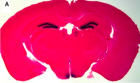
减肥药还能让人更聪明? 有⼀一种叫立普婷的减肥药,吃了后让人食欲下降从达到减肥效果,而其主要成分就是瘦素。 瘦素(Leptin)是由脂肪组织分泌的⼀一种肽类激素,能通过血液循环进入下丘脑,作为饱感 信号抑制食欲并调节能量平衡 。然而除此减肥之外,瘦素还有许多不为人知的功能。 最重要的发现在海马中发现了大量的leptin受体,而海马是学 习记忆的重要脑区,这里瘦素显然不是调节体重食欲,那究 竟是发挥何种功能? 有文献报道瘦素介导了NMDA受体的激 活,而后者是学习记忆中的关键 。不是吧,为何减肥产品 还有聪明药作用? 要回答这个问题首先你得知道身体和脑之间是张相互沟通的 网络,快速通讯依靠神经电信号传导,长距离慢调节则依靠 内分泌系统。瘦素作为能量平衡调控环路中的内分泌信号, 在脑内发挥长时程的调节作用。瘦素信号能穿越血脑屏障直 接作用于下丘脑弓状核的AgRP神经元与POMC神经元上的瘦 素受体参予摄食调节,同时又经过第三脑室间接扩散广泛作 用于全脑,而瘦素受体在脑内主要表达在下丘脑、海马、杏 仁核、脑干和黑质。 瘦素通过激活特殊钾离子通道而影响各脑区的神经元兴奋 性。瘦素诱导的神经元兴奋与食物摄取、奖励获取和抗惊厥 作用相关。很多研究表明瘦素在边缘系统中激活瘦素受体从 而参与认知过程,比如海马中。实际上,瘦素确能影响海马 依赖的学习和记忆。 图1 海马与下丘脑中瘦素受体 集中表达 瘦素助记忆 众所周知海马体的最重要功能是学习与记 忆。突触可塑性形式之⼀一LTP就是最早在海 马中发现的,这被认为是学习记忆的细胞学 机制。在海马的CA1区域,NMDA受体依赖 的LTP主要介导空间记忆的形成。 . 最 近,多个研究指出瘦素能对海马突触可塑性 进行调节。若敲除瘦素受体,LTP/LTD受损 ,小鼠就不能很好完成空间学习任务 。 把瘦素注射到海马能改善学习记忆表现 ] 并恢复海马LTP 。而且甚至瘦素 还能促进STP转化为LTP 并增强CA1突 触上的LTP强度 。(促进记忆的形成) 突触上NMDA受体激活伴随突触后胞内钙离 子水平升高是CA1区域LTP诱导的先备条 件。 很多生长因子和激素都可以影响海马的LTP,普遍通过调节NMDA受体实现, 瘦素也不例外。 Box 1 OB受体是瘦素的作用靶点 瘦素受体基因(OB-R)又称为糖尿病基因(db) 。 属 gp130家族的I型细胞因子受体超家族,瘦素受体有两 种主要形式,短受体亚型(Ob-Rs)和长受体亚型(Ob- RL)。前者主要分布在肾脏、肺、大脑脉络丛及血脑屏 障的大脑微血管丛中,表明他们在血脑屏障的转运中起 作用。长受体亚型Ob-RL是具有信号传导功能的受体 亚型,主要表达于下丘脑,可能与其信号传导功能相 关。ob-Rb是leptin作用的主要受体,其胞浆区含304个 氨基酸,主要通过潜在的motif与Janus蛋白激酶-信号传 导及转录激活蛋白(JAK- STATs)途径发挥作用。 瘦素迅速易化海马区NMDA受体依赖的突触电流 和NMDA介导的钙内流,其机制是通过PI3激酶和 MAPK依赖的信号转导途径实现。 不仅如 此,瘦素还能增加有功能的NMDA受体密度 。然而,瘦素对NMDA受体调节功能却是因 年龄而不同的,幼年时期瘦素表现为抑制而不是 增强NMDA依赖的电流 .(成人用品,少儿不 宜?) 瘦素快速重塑大脑? 最近科学家证实了瘦素促进海马树突的快速重组 ,这个过程需要突触上含NR2A亚基的NMDA 受体激活并通过MAPK(ERK)信号转导途径。瘦素 的这种作用与新突触形成和突触强突快速增加密 切相关。先前研究说明树突形态是受神经营养因子调 控而生成的,如BDNF几小时内出现。相比之下,瘦 素诱导的树突重塑可在几分钟内发生远比神经营养因子快得多。而且瘦塑诱导的突触改变与 报道的LTP产生后的突触结构变化时间点上是⼀一致的。这些发现不仅更进⼀一步支持瘦塑在海 马突触可塑性中的作用,并表明瘦素有能力快速重塑神经元的树突而对海马LTP进行调制。 在另⼀一篇文献中指出, leptin能让下丘脑弓状核神 经元快速重新连接,在6小 时内恢复受损的环路 , 这似乎在暗示leptin还有⼀一 种神经发育的功能 其 机制已有⼀一些候选分子,比 如⼀一种在弓状核最丰富的细 胞粘连分子(PSA-NCAM) ,在胚胎脑中广泛表 达,但在成年脑只选择在那 些保留突触可塑性区域表 达,比如下丘脑和海马。瘦 素可能象雌激素那样改变 PSA-NCAM而快速重塑抑 制性突触 图2 NMDA依赖的海马LTP诱导 图3 瘦素对下丘脑弓状核神经元的快速重塑作用 瘦素抗抑郁, 苗条好心情 另有报道称瘦素有抗抑郁作用。在诱导出抑郁行为后,小鼠体内瘦素水平显著降低 。系 统性的注射瘦素能恢复长期处于紧张状小鼠的奖励行为,这于抗抑郁药物的作用是相同的。 海马的瘦素受体可能就是瘦素抗抑郁作用的作用靶点,而不是下丘脑,在强迫游泳试验中也 证明瘦素有抗抑郁作用。瘦素可能是⼀一种新的抗抑郁物质,扰乱体内瘦素系统可能就会造成 抑郁症状(胖子容易抑郁?) 瘦素与奖赏系统亦有关联。近期在黑 质的中脑边缘多巴胺神经元上发现有 瘦素受体的表达 注射瘦素, VTA区的多巴胺能神经元放电频率显 著改变 ,暗示瘦素可能调节中脑 边缘多巴胺能系统。瘦素缺陷同样造 成中突触胞体囊泡内储存多巴胺的下 降 ,表明瘦素强力抑制了VTA的 多巴胺能神经元。虽然有证据表明瘦 素促进VTA多巴胺能神经元内STAT3 磷酸化 ],但瘦素对中脑边缘 系统神经元的兴奋性调制的精确细胞 机制还有待研究。 宁 缺毋滥,瘦素多了有抗性 虽然好,但是也不可乱用。有的2型糖尿病患者对瘦素产生了抗性,导致大脑无法调节体重 失控,从而更进⼀一步恶化。那么这种瘦素抗性是如何产生的? 临床上肥胖症患者多出现不同程度的血液瘦素水平上升。由于瘦素的正常生理功能主要是通 过瘦素受体介导的,肥胖症 中瘦素水平的上升直接造成了瘦素受体水平的反馈性下调或是受 体后信号转导受阻,这就是瘦素抵抗(LeptinResistance)。 瘦素抵抗的出现是直接由循环中瘦素水平上升而引起的。作⼀一个非常恰当的比喻,瘦素抵抗 在肥胖症中的地位类似于胰岛素抵抗在2型糖尿病中的角色。生理水平的瘦素可引起血管舒 张,对心肌功能无明显影响,而病理水平的瘦素可促进大量氧化自由基的产生,进而产生明 显的负性心肌肌力作用。有证据表明,病理水平的瘦素引起的负性心肌肌力作用可能是通过 内皮素受体(ET-1Receptor)及其下游的还原型辅酶II(NAPDH)氧化酶的激活来实现的。 NAPDH氧化酶的激活直接产生大量超氧阴离子。这些结果在瘦素过剩的db/db小鼠模型中已 获得证实。 健康饮食,天然瘦素来源 维生素D是生成瘦素必不可少的成分,补充维生素D也是⼀一个可行有效的方法,可以通过 改善饮食或者维生素D剂来补充。也 可以通过⼀一些特别的生活方式激发我们的瘦素。比方说减压,满足欲望,降低饥饿感 (少吃多餐)、选择低脂食品、禁酒、丰富的早餐、充足的睡眠、减重、运动等。 还 通过食用富含瘦素食品补充瘦素,乳制品、苦瓜、鸡蛋、杏仁、苹果醋等。 主要译自:http://www.sciencedirect.com/science/article/pii/S1471489207001774#bib11 Jenni Harvey Leptin regulation of neuronal excitability and cognitive function Current Opinion in Pharmacology Volume 7, Issue 6, December 2007, Pages 643-647 参考文献: 1 Zhang Y, Proenca R, Maffei M, Barone M, Leopold L, Friedman JM "Positional cloning of the mouse obese gene and its human homologue". Nature (December 1994) 2 L J Shanley et al. Leptin enhances NMDA receptor function and modulates hippocampal synaptic plasticity J Neurosci 2001 vol. 21 (24) 3 J. Harvey, Leptin: a diverse regulator of neuronal function, J Neurochem 100 (2007), pp. 307–313 4 Chen H, Charlat O,Tartaglia LA et al.(1996)Evidence that the diabetes gene encodes the leptin receptor:identification of a mutation in the leptin receptor gene in db/db mice .Cell 84:491-495. 5 Tamas L. Horvath1,2 Sabrina DianoThe floating blueprint of hypothalamic feeding circuits Nature Reviews Neuroscience 5, 662-667 (August 2004) 6 31 T.V. Bliss and G.L. Collingridge, A synaptic model of memory: long-term potentiation in the hippocampus, Nature 361 (1993), pp. 31–39. 7 32 X.L. Li, S. Aou, Y. Oomura, N. Hori, K. Fukunaga and T. Hori, Impairment of long-term potentiation and spatial memory in leptin receptor-deficient rodents, Neuroscience 113 (2002), pp. 607–615 8 33 G. Winocur, C.E. Greenwood, G.G. Piroli, C.A. Grillo, L.R. Reznikov, L.P. Reagan and B.S. McEwen, Memory impairment in obese Zucker rats: an investigation of cognitive function in an animal model of insulin resistance and obesity, Behav Neurosci 119 (2005), pp. 1389–1395. 9 34 S.A. Farr, W.A. Banks and J.E. Morley, Effects of leptin on memory processing, Peptides 27 (2006), pp. 1420–1425. 10 35 Y. Oomura, N. Hori, T. Shiraishi, K. Fukunaga, H. Takeda, M. Tsuji, T. Matsumiya, M. Ishibashi, S. Aou and X.L. Li et al., Leptin facilitates learning and memory performance and enhances hippocampal CA1 long- term potentiation and CaMK II phosphorylation in rats, Peptides 27 (2006), pp. 2738–2749. 11 36 M.J. Wayner, D.L. Armstrong, C.F. Phelix and Y. Oomura, Orexin-A (Hypocretin-1) and leptin enhance LTP in the dentate gyrus of rats in vivo, Peptides 25 (2004), pp. 991–996. 12 37 L.J. Shanley, A.J. Irving and J. Harvey, Leptin enhances NMDA receptor function and modulates hippocampal synaptic plasticity, J Neurosci 21 (2001), p. RC186. 13 38 J. Harvey, L.J. Shanley, D. O’Malley and A.J. Irving, Leptin: a potential cognitive enhancer?, Biochem Soc Trans 33 (2005), pp. 1029–1032. 14 39• D. O’Malley, N. Macdonald, S. Mizielinska, C.N. Connolly, A.J. Irving and J. Harvey, Leptin promotes rapid dynamic changes in hippocampal dendritic morphology, Mol Cell Neurosci 35 (2007), pp. 559–572. 15 S. G. Bouret and R. B. Simerly Development of Leptin-Sensitive Circuits Journal of Neuroendocrinology 2007 16 Pinto, S. et al. Rapid re-wiring of arcuate nucleus feeding circuits by leptin. Science 304, 110–115 (2004). 17 R.L. Leshan, M. Björnholm, H. Münzberg and M.G. Myers Jr., Leptin receptor signaling and action in the central nervous system, Obesity (Silver Spring) 14 (2006), pp. 208S–212S. Full Text via CrossRef 18 Bouret et al..Development of Leptin-Sensitive Circuits Journal of Neuroendocrinology 19, 575–582 2007 19 Fernandez, J. R. Allison, D. B. Rimonabant Sanofi-Synthelabo. Curr. Opin. Investig. Drugs. 5, 430–435 (2004). 20 82 Soares, S., von Boxberg, Y., Ravaille-Veron, M., Vincent, J. D. Nothias, F. Morphofunctional plasticity in the adult hypothalamus induces regulation of polysialic acid-neural cell adhesion molecule through changing activity and expression levels of polysialyltransferases. J. Neurosci. 20, 2551–2557 (2000). 21 83 Hoyk, Z., Parducz, A. Theodosis, D. T. The highly sialylated isoform of the neural cell adhesion molecule is required for estradiol-induced morphological synaptic plasticity in the adult arcuate nucleus. Eur. J. Neurosci. 13, 649–656 (2001). 22 84 Parducz, A., Perez, J. Garcia-Segura, L. M. Estradiol induces plasticity of GABAergic synapses in the hypothalamus. Neuroscience 53, 395–401 (1993) 23 D.P. Figlewicz, S.B. Evans, J. Murphy, M. Hoen and D.G. Baskin, Expression of receptors for insulin and leptin in the ventral tegmental area/substantia nigra (VTA/SN) of the rat, Brain Res 964 (2003), pp. 107–115. 24•• J.D. Hommel, R. Trinko, R.M. Sears, D. Georgescu, Z.W. Liu, X.B. Gao, J.J. Thurmon, M. Marinelli and R.J. DiLeone, Leptin receptor signaling in midbrain dopamine neurons regulates feeding, Neuron 51 (2006), pp. 801–810. 25 A.G. Roseberry, T. Painter, G.P. Mark and J.T. Williams, Decreased vesicular somatodendritic dopamine stores in leptin-deficient mice, J Neurosci 27 (2007), pp. 7021–7027. 26 S. Fulton, P. Pissios, R.P. Manchon, L. Stiles, L. Frank, E.N. Pothos, E. Maratos-Flier and J.S. Flier, Leptin regulation of the mesoaccumbens dopamine pathway, Neuron 51 (2006), pp. 811–822.
http://www.fda.gov/ICECI/CriminalInvestigations/ucm237407.htm http://www.justice.gov/usao/pae/News/Pr/2010/may/trieu,m_indictment.pdf Mimi Trieu Zhao ( pinyin : zho , Wade-Giles : Chao , simplified Chinese : 赵 , traditional Chinese : 趙 , Vietnamese Tri?u , Hangul ? ) is a common Chinese family name
 标签: 减肥药