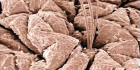
氢气对皮肤移植损伤的保护作用 来自北京协和医院整形外科的王友彬教授课题组的研究,最近在线发表在浙江大学科学 B ( Biomedicine Biotechnology )上, 这是国际上首次关于用氢气治疗皮瓣移植后损伤保护的研究 。皮瓣移植是整形外科重要的手术方式。这应该是整形美容患者的福音。 关于皮瓣,网络上的定义:皮瓣是一具有血液供应的皮肤及其附着的皮下脂肪组织所形成。由于皮瓣自身有血液供应,同时又有皮下脂肪待优点,因而皮瓣移植术的用途不同于游离皮片,主要用于以下几方面: 1 、修复有肌腱、骨、关节 、大血管、神经干等组织裸露的新鲜创面或陈旧性创伤。 2 、器官再造 如鼻、唇、眼睑、眉。 3 、洞穿性缺损的修复 如面颊部洞穿性缺损,除制作衬里外亦常需要具有丰富血运的皮瓣覆盖。 4 、增强局部血运 改善营养状态如放射性溃疡,褥疮等。 皮肤或皮瓣移植手术在整形外科中具有非常重要的地位和作用,但这种手术也会发生一些并发症,最常见的是皮瓣血运障碍。皮瓣出现血液循环障碍,导致皮瓣部分或全部坏死是比较常见的严重的并发症,皮瓣是否出现血循环障碍,从本质上看,就是血液供应是否充分,静脉、淋巴回流是否通畅。也可以发生皮下血肿和感染。大多数皮瓣移植术是为解决患者美观和提高功能问题,一旦失败不仅会给患者带来经济负担,也会给患者和医生带来巨大的精神负担(设想一下,病没有治好,反而增加一个创伤,而且是体表),因此,提高皮瓣移植术成功率,减少并发症是整形外科特别关注的重要问题。但是,目前国际上仍缺乏可靠有明确效果的针对性的理想治疗手段(据说高压氧的效果似乎还不错)。 氢气作为一种理想的无副作用的抗氧化物质,是否可以用于减少皮瓣移植后损伤,值得尝试。 2009 年我在一次学术讨论中提到刚发现氢气具有治疗缺血再灌注损伤的作用,王教授对该信息非常敏感,迅速开展进行这方面的尝试,并很快证明氢气水注射确实可以发挥一定的保护作用,经过随后 3 年不断深入研究,最后形成本研究论文。皮瓣手术在临床上方便用局部给药,将来可以尝试用局部给氢气的方法,不仅可以提高氢气的浓度,而且更容易获得伦理学认可,如果可以证明效果,将具有非常好的应用前景。 hydrogen rich saline on skin flap IR.pdf 论文摘要:皮肤缺血再灌注损伤是一种多因素参与的过程,再整形外科手术经常发生。其机制涉及到缺氧、炎症和氧化损伤。氢气通过选择性抗氧化具有治疗脑缺血的作用。本研究评价了腹腔注射氢气生理盐水对皮瓣移植后缺血再灌注损伤的保护效果。研究选择大鼠腹部皮瓣移植手术为模型,腹腔注射氢气生理盐水为治疗手段,普通生理盐水为对照。手术后连续 5 天治疗后,对皮肤存活面积、局部血流量、活性氧水平和炎症因子水平,心态学分析炎症细胞浸润程度。研究结果发现,氢气生理盐水可以显著提高移植皮瓣存活面积和血流量、氧化损伤和炎症因子等指标明显减轻,局部组织炎症细胞浸润明显减少。研究结果提示,氢气生理盐水注射可以作为减少皮肤缺血再灌注损伤的治疗手段,作用可能和氢气减少移植皮瓣的炎症反应和氧化损伤有关。 Protectiveeffect of hydrogen-rich saline on ischemia/reperfusion in rat skin flap * Ling ZHAO 1 , You-bin WANG †‡2 , Shi-rui QIN 1 ,Xue-mei MA †‡1 , Xue-jun SUN 3 , Ming-lian WANG 1 , Ru-gang ZHONG 1 ( 1 College of Life Science and Bioengineering, BeijingUniversity of Technology, Beijing 100124, China) ( 2 Peking Union Medical College Hospital, Beijing 100032,China) ( 3 Department of Diving Medicine, Faculty of Naval Medicine,Second Military Medical University, Shanghai 200433, China) † E-mail:wy.benz@yahoo.com.cn; xmma@bjut.edu.cn Received Dec. 17, 2012; Revision accepted Apr. 1, 2013;Crosschecked Apr. 8, 2013 Abstract: Objective: Skin damage induced by ischemia/reperfusion (I/R) isa multifactorial process that often occurs in plastic surgery. The mechanismsof I/R injury include hypoxia, inflammation and oxidative damage. Hydrogen (H 2 ) gas has been reported toalleviate cerebral I/R injury by acting as a free radical scavenger. Here, weassessed the protective effect of hydrogen-rich saline on skin flap I/R injury.Methods: Abdominal skin flaps of rats were elevated and ischemia was inducedfor 3 h; subsequently, hydrogen-rich saline (HRS) or physiological saline wasadministered intraperitoneally 10 min before reperfusion. On postoperative d 5,flap survival, blood perfusion, the accumulation of reactive oxygen species andlevels of cytokines were evaluated. Histological examinations were performed toassess inflammatory cell infiltration. Results: Skin flap survival and bloodflow perfusion were improved by HRS relative to the controls. The production ofmalondialdehyde (MDA), an indicator of lipid peroxidation, was markedlyreduced. A multiplex cytokine assay revealed that HRS reduced the elevation inthe levels of inflammatory cytokines, chemokines and growth factors, with theexception of regulated on activation, normal T-cell expressed and secreted(RANTES) growth factor. HRS treatment also reduced inflammatory cellinfiltration induced by I/R injury. Conclusion: Our findings suggest that HRSmitigates I/R injury by decreasing inflammation and, therefore, has the potentialfor application as a therapy for improving skin flap survival.
氢气生理盐水对紫外线 B 皮肤损伤的保护作用 南京医科大学第一附属医院皮肤科最近的研究发现,氢气可以对抗紫外线引起的皮肤损伤,文章发表在 J Biomed Res. (原南京医科大学学报)上。 众所周知,太阳来源的紫外线可以引起皮肤损伤,其中紫外线 B ( UVB )辐射诱导皮肤产生的活性氧( ROS )是导致损伤的重要原因,氢气具有抗 ROS 氧化损伤和抗炎症作用。在本研究中,我们试图证明,富氢生理盐水是否具有保护紫外线辐射皮肤损伤的效果。 选择 C57BL/6 雌性小鼠,经 UVB 辐射照射引起皮肤损伤为疾病模型。给予小鼠腹腔注射富氢生理盐水和富氮生理盐水。显微镜检测伤后皮肤损害。 UVB 辐射后肿瘤坏死因子 α ,白介素( IL ) -1β 和 IL-6 等炎症因子显著升高,组织超氧化物歧化酶降低,丙二醛和一氧化氮的活性升高。富氢生理盐水通过改变这些标志物的水平和改善病理改变。结果表明,富氢生理盐水可能通过减少炎症和氧化应激保护皮肤对抗紫外线辐射损伤。 J Biomed Res. 2012 Sep;26(5):365-71. doi:10.7555/JBR.26.20110037. Epub 2012 Apr 24. Hydrogen-richsaline protects against ultraviolet B radiation injury in rats. Guo Z , Zhou B , Li W , Sun X , Luo D . Source Departmentof Dermatology, the First Affiliated Hospital of Nanjing Medical University,Nanjing, Jiangsu 210029, China; Abstract Exposureof skin to solar ultraviolet (UV) radiation induces photo-damage. Ultraviolet B(UVB) is the major component of UV radiation which induces the production ofreactive oxygen species (ROS) and plays an important role in photo-damage.Hydrogen gas reduces ROS and alleviates inflammation. In this study, we soughtto demonstrate that hydrogen-rich saline has the effect on skin injuries causedby UVB radiation. UVB radiation was irradiated on female C57BL/6 rats to induceskin injury. Hydrogen-rich saline and nitrogen-rich saline were administered torats by intraperitoneal injection. Skin damage was detected by microscope afterinjury. UVB radiation had a significant affection in tumor necrosis factoralpha, interleukin (IL)-1β and IL-6 levels, tissue superoxide dismutase,malondialdehyde and nitric oxide activity. Hydrogen-rich saline had aprotective effect by altering the levels of these markers and relievedmorphological skin injury. Hydrogen-rich saline protected against UVB radiationinjury, possibly by reducing inflammation and oxidative stress. KEYWORDS:
刚看到一篇文章是研究经过皮肤摄取二氧化碳的研究,根据这个思路,氢气、一氧化碳、氧气等是否也可以经过皮肤来摄取,如果设法提高摄取速度,是否可以作为局部疾病治疗的手段。 A Novel System for Transcutaneous Application of Carbon Dioxide Causing an “Artificial Bohr Effect” in the Human Body Background Carbon dioxide (CO 2 ) therapy refers to the transcutaneous administration of CO 2 for therapeutic purposes. This effect has been explained by an increase in the pressure of O 2 in tissues known as the Bohr effect. However, there have been no reports investigating the oxygen dissociation of haemoglobin (Hb) during transcutaneous application of CO 2 in vivo . In this study, we investigate whether the Bohr effect is caused by transcutaneous application of CO2 in human living body. Methods We used a novel system for transcutaneous application of CO 2 using pure CO 2 gas, hydrogel, and a plastic adaptor. The validity of the CO 2 hydrogel was confirmed in vitro using a measuring device for transcutaneous CO 2 absorption using rat skin. Next, we measured the pH change in the human triceps surae muscle during transcutaneous application of CO 2 using phosphorus-31 magnetic resonance spectroscopy ( 31 P-MRS) in vivo. In addition, oxy- and deoxy-Hb concentrations were measured with near-infrared spectroscopy in the human arm with occulted blood flow to investigate O2 dissociation from Hb caused by transcutaneous application of CO 2 . Results The rat skin experiment showed that CO 2 hydrogel enhanced CO 2 gas permeation through the rat skin. The intracellular pH of the triceps surae muscle decreased significantly 10 min. after transcutaneous application of CO 2 . The NIRS data show the oxy-Hb concentration decreased significantly 4 min. after CO 2 application, and deoxy-Hb concentration increased significantly 2 min. after CO 2 application in the CO 2 -applied group compared to the control group. Oxy-Hb concentration significantly decreased while deoxy-Hb concentration significantly increased after transcutaneous CO 2 application. Conclusions Our novel transcutaneous CO 2 application facilitated an O 2 dissociation from Hb in the human body, thus providing evidence of the Bohr effect in vivo .
2011年12月28日发表在《 PLoS ONE 》上的论文表明蚊子咬不咬你可能由你皮肤共生微生物决定。这个研究很有意思,他们找了20-64岁之间的成年男性,实验前禁止饮酒,吃蒜,洋葱等辛辣食物,禁止用香味物质沐浴。给他们穿经过杀菌消毒过的尼龙袜子。采集左脚上的气味物质让蚊子闻,记录对它们的吸引力,之后采集被试左脚底板皮肤上的菌样进行16sRNA分析。研究发现皮肤上面的菌表现高丰度,低多样性的个体更吸引蚊子。高吸引性个体中葡萄球菌Staphylococcus含量是低吸引性个体的2.62倍,而在低吸引性个体身上假单胞菌Pseudomonas的含量是高吸引性个体的3.11倍。贪食菌属Variovoraxspp.和假单胞菌属Pseudomonasspp.与低吸引性群体显著相关。纤毛菌属Leptotrichiaspp.,Delftiaspp.和放线菌Gp3属ActinobacteriaGp3spp.与高吸引性群体显著相关。 只是这个试验有些缺陷,他们用的是脚上皮肤的菌,而且只是给被试穿尼龙袜子但是并没有规定穿什么鞋,鞋可能会影响菌,运动鞋不透气,易出汗,肯定非常臭。出汗多了菌数量应该就多,可能太臭了,氨,硫化氢,吲哚等物质抑制了一些菌的生长导致菌的数量多,但是菌的丰度,也就是菌的种类变少了,因此更加吸引蚊子,所以我推测可能脚越臭越吸引蚊子!这一结果与我们日常生活经验就比较一致了,记得小时候,夏天上床睡觉前妈妈都让洗脚,说这样蚊子不咬,另外去水沟里或泥坑里玩过之后也更招蚊子。 这个结果也算是对“卫生假说”的一个扩展,身体上菌的种类多了,丰富了,身体才能更健康,甚至连蚊子都不咬了,也就很少得疟疾。 如果这一结论是真的的话,将来可以从补充皮肤细菌种类角度开发驱蚊产品,如开发提高皮肤细菌多样性的菌的食物或增殖剂,或者直接喷上与人体皮肤共生的多种细菌。 参考文献: http://dx.doi.org/10.1371/journal.pone.0028991 Composition of Human Skin Microbiota Affects Attractiveness to Malaria Mosquitoes Niels O. Verhulst, Yu Tong Qiu, Hans Beijleveld, Chris Maliepaard, Dan Knights, Stefan Schulz, Donna Berg-Lyons, Christian L. Lauber, Willem Verduijn, Geert W. Haasnoot, Roland Mumm, Harro J. Bouwmeester, Frans H. J. Claas, Marcel Dicke, Joop J. A. van Loon, Willem Takken, Rob Knight, Renate C. Smallegange The African malaria mosquito Anopheles gambiae sensu stricto continues to play an important role in malaria transmission, which is aggravated by its high degree of anthropophily, making it among the foremost vectors of this disease. In the current study we set out to unravel the strong association between this mosquito species and human beings, as it is determined by odorant cues derived from the human skin. Microbial communities on the skin play key roles in the production of human body odour. We demonstrate that the composition of the skin microbiota affects the degree of attractiveness of human beings to this mosquito species. Bacterial plate counts and 16S rRNA sequencing revealed that individuals that are highly attractive to An. gambiae s.s. have a significantly higher abundance, but lower diversity of bacteria on their skin than individuals that are poorly attractive. Bacterial genera that are correlated with the relative degree of attractiveness to mosquitoes were identified. The discovery of the connection between skin microbial populations and attractiveness to mosquitoes may lead to the development of new mosquito attractants and personalized methods for protection against vectors of malaria and other infectious diseases.
原文链接: http://the-scientist.com/2011/11/09/how-skin-tells-time/ 重庆医科大学检验系袁月初译 + 四川大学生物治疗国家重点实验室陈铁林较正。水平有限请大家指正。 How Skin Tells Time 通过肌肤预知时间 The behavior of skin stem cells is regulated by a 24-hour circadian clock. 皮肤干细胞活动受 24 小时生物钟控制 By Ruth Williams | November 9, 2011 Stem cells in the skin, which are responsible for replacing dead skins cells that are continuously sloughed off, follow a daily rhythm that is under the control of a 24-hour circadian cycle, according to a study published today (November 9) in Nature. 11 月 9 日 发表在《 nature 》杂志上的一篇研究表示,皮肤中负责替换持续脱落死细胞的干细胞受 24 小时生物钟控制,呈日节律变化。 “ It was known that in mice proliferation occurs mostly in the night,” said Salvador Aznar-Benitah of the Centre de Regulació Genòmica in Barcelona, Spain, who led the study. “Now that we have added the molecular mechanism we know that this is purely regulated by circadian rhythms.” “众所周知,小鼠(皮肤)增殖大多在晚上,”此项研究的带头人西班牙巴塞罗那 de Regulaci ó Gen ò mica 中心的 Aznar-Benitah 说“我们现在通过对分子机制的研究了解到,这只不过是由 24 小时生物钟所调控。” “ This is a very exciting paper because it links what we knew about signaling molecules …to a much more global regulation—the circadian clock,” said Valentina Greco of Yale University, who was not involved in the study. “It reconciles much of the fragmented information we had,” she said. “这是一篇非常令人兴奋的文章,因为它将(皮肤干细胞的)信号分子与生物钟这个全局调控方式联系起来”未参与研究的耶鲁大学的 Valentina Greco 说“它把很多我们所知的零散的信息联系到了一起” Aznar-Benitah and his colleagues examined the regulation of a known clock protein, Per1, in mouse skin stem cells using a fluorescent protein linked to the clock protein’s promoter. As is the case in other cells of the body, the level of fluorescence oscillated over a 24-hour period. Aznar-Benitah 和他的同事在小鼠皮肤干细胞钟研究了一种时钟蛋白, pre1 的调节。他们将一种荧光蛋白连接到这个时钟蛋白的启动子上。和身体其他细胞中的情况一样,荧光水平 24 小时内变化。 Furthermore, the levels of proliferation-promoting signaling proteins corresponded with the level of the fluorescence—in cells where the fluorescence was bright, proliferation proteins were highly expressed. In dimly fluorescing cells, on the other hand, the expression of such proteins was low. 此外,促增殖信号蛋白水平与荧光水平变化相一致,表现为在细胞中荧光强的部分,促增殖信号蛋白高表达。相反的,在荧光弱的部分,促增殖信号蛋白则低表达。 This suggested that the body’s central clock regulates expression of the signaling proteins in skin, and possibly proliferation. Sure enough, in mice whose skin cells lacked a clock protein called Bmal1, the expression of the signaling molecules was reduced, as was the proliferation of skin stem cells. 这表明了皮肤中信号蛋白受身体的中央生物钟所调控,增殖可能也与此有关。毫无疑问,在皮肤细胞缺乏时钟蛋白 Bmal1 的小鼠中,信号分子表达水平下降,皮肤干细胞增殖水平亦下降。 “ The clock is timing the function of skin cells,” said Aznar-Benitah. “So for example, it is telling the skin that in the morning the main function it has to do is to deal with UV radiation.” Skin stem cells tend to replicate their DNA later in the day to avoid UV-induced mutations, he explained. Indeed, the overall reduction in proliferation in the Bmal1-deficient mice protected them from developing skin cancers. “时钟蛋白控制皮肤细胞的功能” Aznar-Benitah 说“比如,在早晨,他会告诉皮肤,你的主要任务是对付紫外线辐射”他解释道,这样皮肤细胞就将把 DNA 复制推迟以防止紫外线引起其突变。事实上,在 Bmal1 缺乏的小鼠中,整体增殖减少是为了保护他们不发展为皮肤癌。 “ This provides another line of evidence that the clock machinery regulates the protection and activation of the skin cells,” said Cedric Blanpain of the Université Libre de Bruxelles in Belgium, who was not involved in the study. “It has very interesting implications for tumorigenesis,” he added. For example, “if you are jet-lagged and you go into the sun immediately, your cells are much less prepared.” “这篇文章提供了另外一个证据是生物钟肤细胞的保护作用与激活作用” 没有参加研究的比利时 Libre de Bruxelles 大学的 Cedric Blanpain 说“这与肿瘤发生有着很有意思的提示”他补充到,比如“你因为飞机晚点,而后迅速走到太阳下,你的细胞明显就会准备不足。” Although expression of the clock ensures the correct timing of proliferation, many stem cells expressing Bmal1 do not actually replicate. Stem cells in the hair follicle, for example, go through long periods of dormancy where almost no cell proliferation occurs, despite the fact that approximately 50 percent of these cells express the clock proteins. Of these clock-expressing cells, “only 10-20 percent of them actually become active every day,” said Aznar-Benitah. Similarly, only 30-40 percent stem cells outside the follicles replicate each day, though almost all of them express clock proteins, Aznar-Benitah explained. 尽管时钟蛋白的表达确保了增殖的正确时间,但是很多表达 Bmal1 的干细胞实际上并不复制。比如,在头发毛囊里尽管有将近 50% 的细胞表达时钟蛋白,但干细胞将会有很长时间的休眠期以至于几乎没有细胞增殖发生,“在表达时钟蛋白的细胞中,每天只有 10%-20% 被激活” Aznar-Benitah 解释。相同的,在毛囊外尽管他们全部表达时钟蛋白,每天也只有 30%-40% 的干细胞发生复制。 “ The clock is therefore not deterministic but just adds a factor of predisposition to the equation of stem cell activation,” said Aznar-Benitah, “The actual activation depends on the clock and on several other factors—i.e. receiving the activating stimulus.” For skin stem cells, the exact nature of this stimulus is currently unknown. “因此,时钟蛋白不是决定性因素而是干细胞激活反应的一个诱因” Aznar-Benitah 说“真正激活是由时钟蛋白和其它因素控制,如受到激活刺激物刺激。”如皮肤干细胞的确切的刺激物至今还不清楚。 Also unknown is why only some skin stem cells express the clock molecules. “We were really intrigued , and still are, about this finding,” said Aznar-Benitah. “We have some indication that in fact the 50 percent of cells that are not positive might not even be cycling at all, which raises the question of how can a cell be clock-less.” 不清楚的还有为什么只有某些皮肤干细胞表达时钟分子。“我们对此强烈感兴趣,并且没有持续寻找原因” Aznar-Benitah 说“我们发现了一些现象表明,事实上,那 50% 不活化(时钟蛋白不活化)的(毛囊)细胞可能甚至都不参与细胞循环 及细胞分裂,这就产生了一个问题:细胞怎么可以缺少生物钟控制呢?” P. Janich et al., “The circadian molecular clock creates epidermal stem cell heterogeneity,” Nature, doi:10.1038/nature10649, 2011.
据《科学·内幕》( ScienceInsider )2011年9月12日报道,美国芝加哥大学的另外一个实验室可能受到污染,因为两年前曾经有一位研究者在此因感染瘟疫随后死亡。2011年8月,在同样的实验室所在地,有研究人员因为皮肤感染而住院,但是导致感染的是一种该实验室正在研究的常见的蜡样芽胞杆菌(Bacillus cereus)。 据芝加哥大学透露,从事蜡样芽胞杆菌这个项目研究的是微生物科学家Olaf Schneewind。2011年8月27日,她被送进医院,在接受手术治疗和抗生素药物治疗之后出院。在她的实验室,蜡样芽胞杆菌的研究是在生物安全性二级水平的条件下进行的,这是属于4个生物安全性水平中较低的一种。芝加哥大学在2011年9月初已经暂停该研究区域,进行清洁处理,以预防类似事件再次发生,但是期待实验室将在一周之后重新开放启用。 住院治疗的研究者很可能通过伤口暴露而被感染,但是芝加哥大学对此展开调查,到底她是否是在实验室里而被感染。只要按章操作,随后及时洗手,蜡样芽胞杆菌是不会传染的,但为了避免传染的危险,家人和同事都应该回避,有些则提供了抗生素来进行预防。两年前,在生命科学中心同一区域工作的一个研究员即遗传学家Malcolm Casadaban, 也是Schneewind研究项目的副主持人(co-principal investigator),受到一种被认为不会感染健康成人的鼠疫耶氏菌(Yersinia pestis)的感染而最终死亡。根据一份美国疾病控制和预防中心的发病率和死亡率周报(Mortality and Morbidity Weekly Report,简称MMWR)报道,Casadaban可能生病,因为他已经有血色素沉着即体内铁贮积过多所致,而且在Casadaban体内,因为铁过量可能会引起毒副作用更强。报告称Casadaban使用的手套也不一致,可能是在使用手套过程中,通过暴露的真皮感染而最终导致死亡,蜡样芽胞杆菌的感染是否也是具有相同的传播途径而导致微生物科学家Olaf Schneewind住院?芝加哥大学的公共健康部门已走访了校园,并对于实验室的安全程序进行评估。案件既不涉及到一种选择试剂,也不涉及到疾病预防控制中心列入黑名单的生物攻击潜在试剂中的某一种病原体。虽然蜡样芽胞杆菌属于名单内容之一,但是对于Casadaban的研究例外。这件事情的出现,等于给所有从事生物制剂研究的机构提了个醒,在研究过程中从规范操作到使用药品的妥善管理,丝毫不可粗枝大叶。 部分英文择选如下: Two years ago, a researcher who worked in the same area in the Cummings Life Science Center, geneticist Malcolm Casadaban, a co-principal investigator with Schneewind, died after becoming infected with a weakened strain of the Yersinia pestis bacterium that was not thought to infect healthy adults. According to a report in the Centers for Disease Control and Prevention's Mortality and Morbidity Weekly Report , Casadaban may have become sick because he had hemochromatosis, or an overload of iron in the body. The Y. pestis strain had been weakened by making it less able to acquire iron, and the excess iron in Casadaban's body might have allowed it to be become more virulent, the MMWR report says. Neither case involved a select agent—a pathogen on CDC's list of potential agents in a biological attack. (Although Y. pestis is on the list, the strain Casadaban studied was excluded.) But Schneewind also directs the Great Lakes Regional Center of Excellence for Biodefense and Emerging Infectious Diseases Research , a consortium funded by the National Institute of Allergy and Infectious Diseases (NIAID) to study select agents and natural threats. The center does some of its work at a major biosafety level-3 lab on the campus of Argonne National Laboratory, one of a dozen such regional biocontainment labs built partly with NIAID funding after the 2001 anthrax attacks.
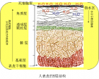
(文中引用资料尽可能采用可靠来源,为不影响阅读以超链接形式引用,有意核实者请查阅原材料。 文中观点为个人观点,不代表任何利益。) Figure Diseases linked to nanoparticles from different pathways of exposure. 图片引自: http://www.i-sis.org.uk/nanotoxicityInRegulatoryVacuum.php copyrighted. (only for personal use.) 据调查, 由于澳大利亚全年大部分时间阳光充足,紫外线强烈,人们又热衷户外运动,是全世界皮肤癌发病率最高的国家之一,也是使用防晒霜最广泛的地区之一。 最近全国气温飙升 ,很多地区也达到四十度左右,也是阳光明媚,夏日炎炎。不知用防晒霜的人多不多?印象中我自己以前极少用。去年用过几天,后来就不用了。目前,权威部门或医生通常还会建议使用防晒霜。但随着研究深入,这个问题越来越值得商榷。 普通民众使用防晒霜应该很少关心其作用原理,想当然地以为防晒霜就是防晒的。选择时估计也就关心哪个闻起来香,一般不会关心会有什么健康影响。 不过,学术界 近年来越来越关注 防晒霜的健康影响 。主要原因是由于流行的防晒霜很多采用了纳米颗粒来反射紫外线。目前 很多类型的防晒霜 已经使用20~30纳米(nm)大小的氧化锌(ZnO)或二氧化钛(TiO 2 )颗粒。这样大小的微粒的尺寸是可以和DNA相比较的。我们知道DNA通常为2nm左右,而人的红细胞有7000nm。从尺度比较就可知道20nm左右的颗粒更可能和DNA分子作用,其特性也是较难全面了解的,其生物特性也较难以验证。这也是目前科学研究的一个分支之一。 图 皮肤模型。源自Shier, D., Butler, J. Lewis, R. in Hole's Human Anatomy and Physiology 8th Edn 160183 (McGraw Hill, 1999 ). and http://www.nature.com/nature/journal/v445/n7130/full/nature05664.html#B79 COPYRIGHTED. (only for personal use.) 研究已经证明生物吸入或注射 纳米颗粒会引起明显且严重的健康变化 。 更严重的是,几十纳米大小的微粒很容易进入细胞内部。这是引起关注的主要原因之一。以前使用的防晒霜里的成分大多尺寸较大,在微米量级,几乎不可能穿过细胞壁进入细胞内部。微米量级的微粒性能和宏观材料基本一致。如微米级的氧化锌等呈白色,也是以前的防晒霜涂在身上发白的原因。而一旦微粒在纳米量级(100nm)会变为透明,所以现在的防晒霜抹上一般就是无色的了。但其作用也更难以预料。 目前的研究结果虽然提出 很多质疑 ,但尚未确实证明防晒霜对人体的健康影响。权威部门会要求生产厂商在产品封装上 标明使用纳米颗粒 ,基本是把选择权留给消费者。 不过,我想大部分相关研究人员会持保留意见,希望消费者在没有明确试验结论之前, 尽量减少使用量, 对自己健康负责。人体是个复杂的系统,任何微妙改变都可能引发一系列严重问题。 不要小瞧防晒霜可能带来的健康影响。 当阳光照耀防晒霜的时候,当你沉浸在温暖的阳光中享受浪漫的时刻,你身上正发生着全世界数量庞大的科学家群体,日以继夜,夙兴夜寐,而无法搞清楚的巨复杂的化学、物理、生物复合的变化过程 更多详细专业信息可以参考一些开放的网络信息,如 http://www.jltp.uiuc.edu/archives/Abramowitz.pdf 。 图片引自 http://www.nextnature.net/2009/10/nanoparticles-in-sunscreen-damage-microbes/
 标签: 皮肤
![[转载]护肤](http://image.sciencenet.cn/album/201205/28/125423tjcj7jxzaoacjjro.jpg.thumb.jpg)