博文
以甲烷为食的细菌将温室气体转化为燃料
 精选
精选
||
以甲烷为食的细菌将温室气体转化为燃料
诸平
据美国西北大学(Northwestern University)2022年3月17日报道,以甲烷为食的细菌将温室气体转化为燃料(Methane-eating bacteria convert greenhouse gas to fuel)。
甲烷营养菌(Methanotrophic bacteria)每年消耗3000万吨甲烷,它们将这种强效温室气体转化为可用燃料的天然能力吸引了研究人员。然而,我们对复杂反应是如何发生的了解甚少,限制了我们利用这双重利益的能力。通过研究细菌用来催化反应的酶,美国西北大学的一个研究小组现在发现了可能推动这一过程的关键结构。相关研究结果于2022年3月18日将在《科学》(Science)杂志网站发表——Christopher W. Koo, Frank J. Tucci, Yuan He, Amy C. Rosenzweig. Recovery of particulate methane monooxygenase structure and activity in a lipid bilayer. Science, Published Date: 17 Mar 2022. 375(6586): 1287-1291. DOI: 10.1126/science.abm3282. http://www.science.org/doi/10.1126/science.abm3282.这项研究结果最终可能会导致将甲烷气体(methane gas)转化为甲醇的人造生物催化剂的开发。
这篇论文的通讯作者、西北大学的艾米·罗森茨韦格(Amy Rosenzweig)说:“甲烷有一个非常强的化学键,所以有一种酶能做到这一点是非常了不起的。如果我们不确切地了解酶是如何执行这种困难的化学反应的,我们就无法为生物技术应用设计和优化它。”
艾米·罗森茨韦格是西北大学温伯格艺术与科学学院(Northwestern's Weinberg College of Arts and Sciences)温伯格家族生命科学杰出教授(Weinberg Family Distinguished Professor of Life Sciences),在那里她担任分子生物科学和化学的职务。
这种酶被称为颗粒状甲烷单加氧酶(particulate methane monooxygenase简称pMMO),是一种特别难以研究的蛋白质,因为它嵌入在细菌的细胞膜(cell membrane)中。
通常,当研究人员研究这些甲烷营养菌时,他们会使用一种苛刻的过程,用洗涤剂溶液将蛋白质从细胞膜上剥离。虽然这一过程有效地分离了酶,但它也杀死了所有的酶活性,并限制了研究人员能够收集的信息,就像监测没有心跳的心脏一样。
在这项研究中,该团队完全使用了一种新技术。艾米·罗森茨韦格实验室的博士研究生、此项研究的第一作者克里斯托弗·古(Christopher Koo)想知道,如果把酶放回与自然环境相似的膜中,他们是否能学到新的东西。克里斯托弗·古利用细菌的脂质在一种名为纳米圆盘(nanodisc)的保护性颗粒内形成一层膜,然后将酶嵌入到这层膜中。
克里斯托弗·古说:“通过在纳米圆盘内重建酶的自然环境,我们能够恢复酶的活性。然后,我们能够使用结构技术在原子水平上确定脂质双分子层是如何恢复活性的。在此过程中,我们发现了酶中铜位点的完整排列,甲烷氧化很可能发生在这里。”
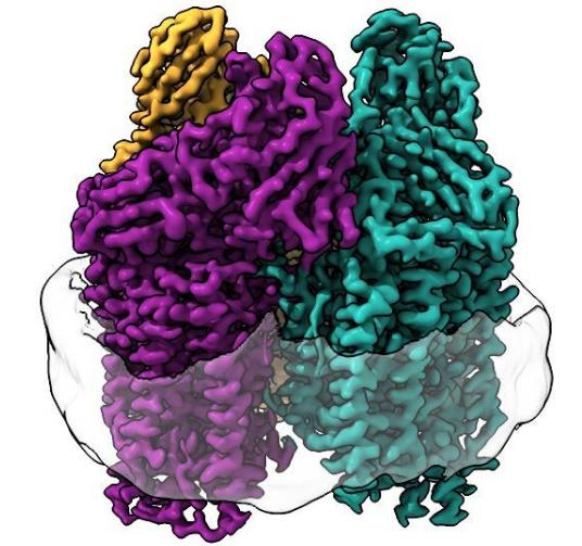
研究人员使用了低温电子显微镜(cryo-electron microscopy简称cryo-EM),这是一种非常适合膜蛋白的技术,因为在整个实验过程中脂膜环境没有受到干扰。这使得他们第一次以高分辨率看到活性酶的原子结构。上图就是由西北大学提供的低温电镜显示出了蛋白质膜中前所未见的结构。
艾米·罗森茨韦格说:“由于最近低温电子显微镜的‘分辨率革命’,我们能够看到原子结构的细节。我们所看到的完全改变了我们对这种酶活性部位的看法。”
艾米·罗森茨韦格说,低温电镜结构为回答不断堆积的问题提供了一个新的起点。甲烷是如何到达酶活性部位的?或者甲醇是如何从酶中出来的?活性部位的铜是如何进行化学反应的?接下来,该团队计划使用一种称为低温电子断层扫描(cryo-electron tomography简称cryo-ET)的前沿成像技术直接研究细菌细胞中的酶。
如果成功,研究人员将能够准确地看到酶在细胞膜中的排列方式,确定它在真正的自然环境中是如何工作的,并了解酶周围的其他蛋白质是否与它相互作用。这些发现将为工程师提供一个关键的缺失环节。
艾米·罗森茨韦格说:“如果你想优化这种酶,将其插入生物制造过程中,或者消耗除甲烷以外的污染物,那么我们需要知道它在自然环境中的样子,以及甲烷在哪里绑定。你可以用细菌和一种改造过的酶从压裂现场(fracking sites)收集甲烷,或者清理石油泄漏。”
上述介绍,仅供参考。欲了解更多信息,敬请注意浏览原文或者相关报道。
消耗甲烷的细菌可能会成为未来的燃料(Methane-consuming bacteria could be the future of fuel)
Getting the environment just right
Particulate methane monooxygenase (pMMO) is a key enzyme for the metabolism of methane by aerobic, methanotropic bacteria. There is considerable interest in understanding this reaction, both as a fundamental biogeochemical transformation and for potential biotechnological applications, but progress has been hampered by inactivation of this membrane-bound metalloenzyme upon purification. Koo et al. reconstituted natively produced pMMO in lipid nanodisks and found that restoration of the bilayer environment also restored activity of the enzyme. A series of cryo–electron microscopy structures revealed previously missing regions of the enzyme, including conserved residues that coordinate a metal ion near the inner pore of the trimeric complex in a site not seen in previous structures. Lipids play an important role in stabilizing various parts of the structure, and the recovery of activity in these samples is consistent with the newly observed metal-binding site being involved in methane oxidation. —MAF
Bacterial methane oxidation using the enzyme particulate methane monooxygenase (pMMO) contributes to the removal of environmental methane, a potent greenhouse gas. Crystal structures determined using inactive, detergent-solubilized pMMO lack several conserved regions neighboring the proposed active site. We show that reconstituting pMMO in nanodiscs with lipids extracted from the native organism restores methane oxidation activity. Multiple nanodisc-embedded pMMO structures determined by cryo–electron microscopy to 2.14- to 2.46-angstrom resolution reveal the structure of pMMO in a lipid environment. The resulting model includes stabilizing lipids, regions of the PmoA and PmoC subunits not observed in prior structures, and a previously undetected copper-binding site in the PmoC subunit with an adjacent hydrophobic cavity. These structures provide a revised framework for understanding and engineering pMMO function.
https://m.sciencenet.cn/blog-212210-1330044.html
上一篇:可堆叠的“全息砖”可以制作巨大的3D图像
下一篇:美中日物理学家首次在二维材料中发现了强电子相关性的直接证据