博文
超清版神经突触结构
|||
http://www.sciencemag.org/content/344/6187/1023
感谢Silvio Rizzoli教授提供全文
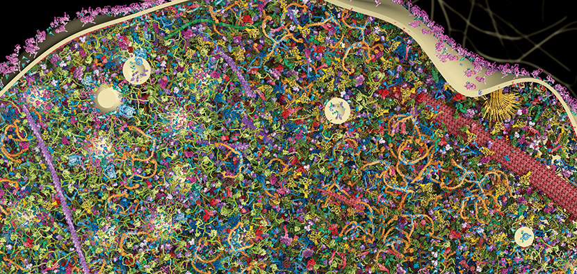
不得不说,这是科学和技术的完美结合,欧洲得大脑图计划只是对大脑得神经纤维联系进行准确描绘和功能模拟,似乎并没有包括对一个神经细胞或某一部分进行高分辨率三维重建,不过这期《科学》封面文章就是一幅神经突触的超高分辨率图,这个图是结合多种技术获得模拟图,这是目前多种显微技术的完美结合。

这一研究首先使用量化免疫杂交和质谱分析决定蛋白质的类型和浓度,电子显微镜检测细胞器的数量、大小和位置,超分辨率荧光显微镜定位蛋白质。使用这些数据,这些科学家制作出一个神经突触的三维模型,模型能在原子层面上显示30万种蛋白质,研究还发现,在突触囊泡循环的同一步骤,这些蛋白质的数量比较接近,但在不同步骤如在囊泡融合和泡吐之间,这些蛋白的拷贝数从150到2万,相差可达3个数量级。
在这个研究中,也很好地演示了形态学和量化检测指标各自的特征,并将两者进行了很好的取长补短。量化研究方法,如果采用蛋白电泳杂交,只能半定量蛋白数量,其实是将不同蛋白分离,然后用免疫学(抗原抗体反应)方法半定量目标蛋白,质谱则能够更准确地定量分析,电泳结合质谱分析,大概是对复杂生物样品蛋白质浓度分析的最理想手段了。不过这种已经达到极致的定量技术无法提供任何定位信息,或者说完全不知道这些蛋白都分布那些部位,相对浓度如何(不可能是均匀分布),这就需要免疫荧光技术上场了,免疫荧光是通过免疫学(仍然是抗原抗体反应)技术,将目标蛋白在显微层面进行定位,当然也可以根据荧光相对强度,更低程度(比电泳更不准确)定量。不过免疫荧光要结合显微镜技术,如果是光学显微镜,那么你只能获得光学显微镜级别的图像,如果是电子显微镜,那么可以将分辨率有效提高。最后是计算机模拟技术,对上述这种海量的数据进行整合,就必须采用计算机技术,甚至需要超级计算机的运算能力。这也是欧洲脑图计划所面临的技术瓶颈之一。如果欧洲大脑图计划类似人口普查,而这个技术则是对一个人进行大体进行更细致描述了。
牛角尖:这个高精尖技术并没有将非蛋白类成分包含在内,显然组成细胞的成分还有水、各种离子和小分子有机物,也有脂和糖类大分子。如果能将这些信息整合在一起,或者能更加准确地反映突触的功能细节。不过目前技术看,这些工作存在更大挑战性。不过目标在前,勇者胜,科学研究的特点是不怕做不到,就怕想不到。
This,in all its molecular complexity, is what thebulging end of a single neuron looks like. A whopping 300,000 proteins cometogether to form the structure, which is less than a micrometer wide, hundredsof times smaller than a grain of sand. This particular synapse is from a ratbrain. It’s where chemical signals called neurotransmitters are released intothe space between neurons to pass messages from cell to cell. To create a 3Dmolecular model of the structure, researchers first isolated the synapses ofrat neurons and turned to classic biochemistry to identify and quantify themolecules present at every stage of the neurotransmitter release cycle. Then,they used microscopy to pinpoint the location of each protein. Someproteins—like the red patches of SNAP25 visible in the video at 0:14—aid in therelease of vesicles, tiny spheres full of neurotransmitters. Others—like thegreen, purple, and red rods at 0:45—help the synapse maintain its overallstructure. Different proteins surround vesicles when they’re inside thesynapse—the circles scattered throughout the structure at 0:56—than when thevesicles are forming at the edge of the synapse—as shown at 2:08. Researcherscan use the model, described online today in Science, tobetter understand how neurons function and what goes wrong in brain disorders.
https://m.sciencenet.cn/blog-41174-798841.html
上一篇:小保方同意撤回《自然》STAP细胞论文
下一篇:9名科学家分享2014年Kavli科学奖