博文
X射线成像技术在不同研究领域的典型应用
|
Article title: Methodology development and application of X-ray imaging beamline at SSRF
上海光源(SSRF)X射线成像线站实验方法研究和应用研究最新进展
DOI: https://doi.org/10.1007/s41365-020-00805-7
One sentence summary:
一句话概要:
This study further explores the potentials of X-ray imaging and contributes to the development of numerous new applications.
本报告将深入探讨X射线成像技术所蕴含的巨大潜力,为更多全新应用的研发提供助力
The Novelty (What)
创新性(主要内容)
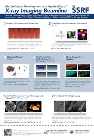
This study reports the latest developments of X-ray imaging methodology and new applications of the X-ray imaging and biomedical application beamline (BL13W1) in the past 5 years at the Shanghai Synchrotron Radiation Facility (SSRF). By equipping it with several sets of X-ray imaging detectors of different pixel sizes (0.19 – 24 mm), the beamline (photon energy range of 8–72.5 keV) can realize X-ray micro-computed tomography (X-ray micro-CT) and X-ray in-line phase-contrast imaging. Based on these two basic methods, several x-ray imaging methods and image processing techniques were developed. Firstly, in vivo dynamic micro-CT experiments with living insects were successfully conducted in 0.5 s (sampling rate of 2 Hz, 2 tomograms/s) with a monochromatic beam and 40 ms (sampling rate of 25 Hz, 25 tomograms/s) with a white beam. Next, move contrast X-ray imaging (MCXI) was developed whereby the blood flow and moving tissues in raw images became distinguishable. Furthermore, the attempt to use X-ray speckle-tracking imaging with twice exposures to eliminate the edge enhancement effect was successfully developed. A high-precision quantification method via X-ray micro-CT to measure complex three-dimensional (3D) blood vessels was also developed. Besides, there was the development of an X-ray nano-CT with 100 nm spatial resolution too. By combining the X-ray micro-CT imaging method with other contrast mechanisms, three-dimensional X-ray diffraction (3DXRD) microscopy, small-angle X-ray scattering CT (SAXS-CT), and X-ray fluorescence CT (XFCT) were developed. These x-ray imaging methods and image processing techniques have been proven to bring the studies of material science, biomedicine, paleontology, physics, chemistry, and environmental science to a whole new level. With continuous research and development, it is clear that more experimental methods and techniques will be developed and provided to users to achieve more high-level applications.
本报告论述了在过去五年中,上海光源 (Shanghai Synchrotron Radiation Facility, SSRF)X射线成像及生物医学应用光束线站 (BL13W1)在X射线成像实验方法学研究及应用研究的最新进展。该光束线站配置了多套X射线成像探测器 (像素尺寸0.19 – 24 mm), 光子能量范围8–72.5 keV,可实现X射线计算机断层扫描(X射线显微CT)和X射线同轴相衬成像。在此基础上发展了几种新的X射线成像方法以及图像处理技术。基于单色光的活体昆虫动态显微CT实验在0.5秒内完成 (采样率2Hz,2套CT数据/秒);基于弯铁白光的活体昆虫动态显微CT实验在40毫秒内完成 (采样率25Hz,25套CT数据/秒)。运动衬度X射线成像(Move contrast X-ray imaging, MCXI)技术被开发出来,原始图像中的血液流动以及组织运动情况因此得以清晰显现。X射线散斑跟踪成像两次曝光技术被采用,成功消除了边缘增强效应。基于X射线显微CT实现对复杂血管进行高精度的三维结构定量表征的图像处理技术被成功开发。100纳米空间分辨率的X射线纳米CT也被开发出来。此外,我们把X射线显微CT成像技术与其他相衬机制相结合,开发出三维X射线衍射 (Three-Dimensional X-ray Diffraction, 3DXRD)显微技术、X射线小角散射CT(Small-Angle X-ray Scattering CT ,SAXS-CT)技术以及X射线荧光CT(X-ray Fluorescence CT, XFCT)技术。以上X射线成像方法以及图像处理技术均已被证实可将材料科学、生物医学、古生物学、物理学、化学以及环境科学提升至更高研究水平。通过不断研究和开发,会有更多实验方法和技术被开发出来并推向市场,为用户带来更高水平的应用。
The Background (Why)
研究背景(主要原因)
With rapid technological advancement, there is a demand for research and development sectors to keep expanding and evolving. As a result, researchers are constantly setting up facilities, developing new research methodologies, and exploring new applications of existing equipment. X-ray was discovered more than a century ago. With an in-depth understanding of its properties, new applications could be derived by including additional features or integrating different tools. This study has reported that, within five years, multiples X-ray imaging methodologies and applications have been newly developed at the SSRF. Up to this point, the development has managed to benefit research in various fields, including material science, biomedicine, paleontology, physics, chemistry, and environmental science. Thus, more advanced studies can be conducted to enhance the technology with further improvement and innovation in developing X-ray imaging methods and techniques.
随着技术的快速提升,科研与研发事业将不断扩大并持续演进。在此背景下,科研人员需要安装设备、开发全新研究方法、探索已部署设备的全新用途。X射线发现已超过一个世纪,随着业界对其属性了解得愈发深入,通过添加更多功能或与更多不同工具实现集成,越来越多的全新应用被引入进来。本篇论文将重点介绍在过去5年中,上海光源(SSRF)开发出的多项X射线成像技术和应用。目前为止,这些技术和应用已经惠及诸多领域,如材料科学、生物医学、古生物学、物理学、化学以及环境科学等。随着X射线成像方法以及技术的不断提升和创新,越来越多的研究将得以展开。
The SDG impact (Big Why)
SDG影响力(研究意义)
Innovation and technological progress are key to finding lasting solutions to both economic and environmental challenges. Hence, scientific research and development play a crucial role in advancing technology and developing new methods to benefit our everyday routines. The X-ray imaging methodologies and techniques reported by this study have proven their respective potential in various fields of study. Since one of the United Nations’ main interests is to enhance scientific research, this study sets the path to realizing the objectives of UNSDG 9: Industries, Innovation & Infrastructure.
面对经济和环境挑战,最为有效的方法是实现技术创新和进步,从一定意义上说,科学研究在尖端技术开发与寻找全新方法方面扮演着极其重要的角色,并最终惠及大众生活。本报告中所论及的X射线成像方法及技术蕴含巨大潜力,这已在各个领域的研究中获得证实。联合国的任务之一,就是推动并提升科学研究水平,本报告将为联合国可持续发展17个目标中的第9个目标(UNSDG 9),即工业、创新和基础设施的实现提供全新路径。
https://m.sciencenet.cn/blog-3474219-1365971.html
上一篇:一套新开发的可应用于核物理基础研究的探测系统
下一篇:通过动态几何信息交换提高液基探测器模拟精度